|
|
|
|
|
|||||||||||
|
Electronic Flora of South Australia Species Fact Sheet
Phylum Rhodophyta – Family Delesseriaceae
Selected citations: Millar & Kraft 1993: 48. Silva et al. 1996: 464.
Thallus (Fig. 29E) medium red-brown, decumbent to more-or-less erect, 3–8 cm high, densely tufted with complanately branched primary and main blades 3–5 mm broad, bearing marginally lateral blades (1–) 2–4 mm broad with slender, compressed to terete, basal constrictions 200–500 µm broad; blades linear to gently tapering, flat, apices acute to rounded, margins with short, blunt, dentations; all blades with a central midrib and relatively faint, paired, lateral veins (Fig. 30A). Attachment by small discoid holdfasts bearing 1–3 fronds, often with a stoloniferous base giving rise to further erect fronds; epiphytic, epizoic (e.g. on bryozoans) or epilithic. Structure. Apical cell conical, segmenting rapidly (Fig. 29F) to an axial row (which soon becomes corticated) and lateral and transverse pericentral cells, the lateral cells producing second-order rows reaching the blade margin and from which third-order cells or short rows are cut off abaxially and adaxially in an irregular manner, giving a monostromatic blade (apart from the veins - Fig. 29G) of compact, isodiametric, irregular cells 20–30 µm across in surface view; lateral veins with central primary cells and a cortex 1 (–2) cells thick; transverse intercalary divisions occur in the axial cells so that the second-order rows become separated by 2–5 axial cells as they develop into the lateral paired veins, with further lateral divisions of the daughter axial cells and the second- and third-order cells forming the expanded blade. The margins of the blade bear short dentations 30–50 (–200) µm long, the larger ones corresponding to the vein endings and occasionally developing as lateral blades. Mature cells multinucleate; rhodoplasts discoid.
Reproduction: Gametophytes dioecious. Procarps scattered on both sides of blades, probably with a 4-celled carpogonial branch and 2 sterile groups. Carposporophytes with a small basal fusion cell and chains of ovoid carposporangia 15–30 µm in diameter. Cystocarps (Fig. 30B) 300–700 µm across, few per blade, protruding mainly on one side of the blade, ostiolate; pericarp 90–140 µm and 5–8 cells thick, thicker around the ostiole (Fig. 30C). Spermatangial sori (Fig. 30D) on both sides of blades between midrib and margins, covering the lateral veins, with each primary cell covered by 8–10 cortical cells each of which produces 2–4 spermatangia. Tetrasporangial sori (Fig. 30E) scattered, often close to margins, with tetrasporangia in 2 layers (Fig. 30F), cut off from inner cortical cells and covered with a layer of small outer cortical cells, subspherical and 30–50 µm in diameter.
Type from Gellibrand Light, northern Port Phillip Bay, Vic.; holotype in MELU, A35185, with isotypes.
Selected specimens: Port Noarlunga, S. Aust., upper sublittoral on reef (Wollaston, 26.x.1965; AD, A29711). Gloucester Reserve, Williamstown, Vic. 2–4 m deep (Kraft & Saunders, 6.ii.1995; MELU). Point Lonsdale, Vic., on sponge 1–4 m deep (Kraft & Saunders, 27.ii.1992; MELU, K8946). Crawfish Rock, Westernport Bay, Vic. (Watson, Nov. 1968; AD, A32138), 3–6 m deep (Watson, 15.ix.1965; AD, A32823), 6 m deep (Watson, 26.iv.1969; AD, A34369), 15 m deep (Watson, 5.iv.1970; AD, A35833), 5–10 m deep (Watson, 29.viii.1971; AD, A39370), 6 m deep (Shepherd, 4.ii.1971; AD, A37900), 10 m deep (Shepherd, 4.ii.1971; AD, A37940), 6 m deep (Watson, 16.i.1974; AD, A44854), and 10 m deep (Watson, 28.v.1974; AD, A45432).
Distribution: Hamelin Bay, W. Aust., Port Noarlunga, S. Aust., Port Phillip Bay and Westernport Bay, Vic., and N.S.W. and Norfolk and Lord Howe Is.
Taxonomic notes: P. australasica is a common alga in Port Phillip Bay and at Crawfish Rock, Westernport Bay, Vic., present throughout the year, and is probably more generally distributed in suitable localities than the other scattered collections indicate.
References:
MILLAR, A.J.K. & KRAFT, G.T. (1993). Catalogue of marine and freshwater Red Algae (Rhodophyta) of New South Wales, including Lord Howe Island, South-western Pacific. Aust. Syst. Bot. 6, 1–90.
MILLAR, A.J.K. (1990). Marine Red Algae of the Coffs Harbour Region, northern New South Wales. Aust. Syst. Bot. 3, 293–593.
MILLAR, A.J.K. (1999). Marine benthic algae of Norfolk Island, South Pacific. Aust. Syst. Bot. 12, 479–547.
SILVA, P.C., BASSON, P.W. & MOE, R.L. (1996). Catalogue of the Benthic Marine Algae of the Indian Ocean. (Univ. California Press: Berkeley.)
The Marine Benthic Flora of Southern Australia Part IIID complete list of references.
Publication:
Womersley, H.B.S. (24 February, 2003)
The Marine Benthic Flora of Southern Australia
Rhodophyta. Part IIID. Ceramiales – Delesseriaceae, Sarcomeniaceae, Rhodomelaceae
Reproduced with permission from The Marine Benthic Flora of Southern Australia Part IIID 2003, by H.B.S. Womersley. Australian Biological Resources Study, Canberra. Copyright Commonwealth of Australia.
Illustrations in Womersley Part IIIA, 2003: FIGS 29 E–G, 30.
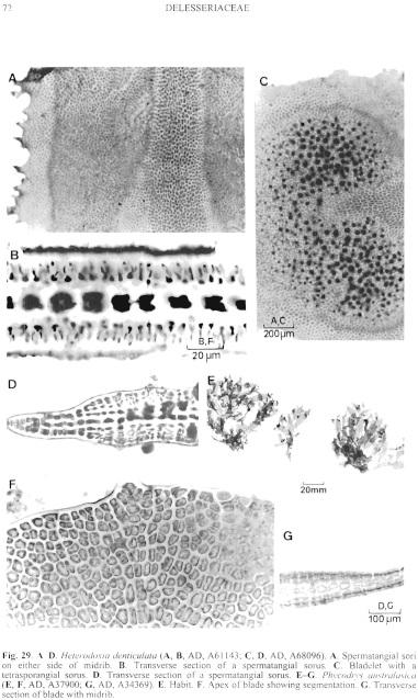
Figure 29 enlarge
Fig. 29. A–D. Heterodoxia denticulata (A, B, AD, A61143; C, D, AD, A68096). A. Spermatangial sori on either side of midrib. B. Transverse section of a spermatangial sorus. C. Bladelet with a tetrasporangial sorus. D. Transverse section of a spermatangial sorus. E–G. Phycodrys australasica (E, F, AD, A37900; G, AD, A34369). E. Habit. F. Apex of blade showing segmentation. G. Transverse section of blade with midrib.
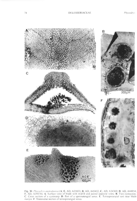
Figure 30 enlarge
Fig. 30. Phycodrys australasica (A, E, AD, A35833; B, AD, A45432; C, AD, A34369; D, AD, A44854; F, AD, A39370). A. Surface view of blade with midrib and paired opposite veins. B. Two cystocarps. C. Cross section of a cystocarp. D. Part of a spermatangial sorus. E. Tetrasporangial sori near blade margin. F. Transverse section of tetrasporangial sorus.

|
Email Contact: State Herbarium of South Australia |

|


