|
|
|
|
|
|||||||||||
|
Electronic Flora of South Australia Species Fact Sheet
Phylum Rhodophyta – Family Delesseriaceae
Selected citations: Huisman & Walker 1990: 429. Millar & Kraft 1993: 45. Silva et al. 1996: 447. Wynne 1984a: 141, figs 16–26.
Synonyms
Delesseria spathulata Sonder 1845: 57; 1848: 194; 1881: 105. J. Agardh 1852: 698; 1872: 58. Harvey 1855a: 548; 1863, synop.: xxxi. Kützing 1869: 5, pl. 12c-e.
Hypoglossum spathulatum (Sonder) Kützing 1849: 877.
Delesseria tasmanica Mueller ex Harvey 1859b: 311, pl. 190B; 1863, synop.: xxxi. J. Agardh 1872: 58; 1876: 494. Kützing 1869: 4, pl. 11a, b. Reinbold 1897: 14. Sonder 1880: 24. Tisdall 1898: 509.
Apoglossum tasmanicum (Mueller) J. Agardh 1898: 194. De Toni 1900: 702. Guiler 1952: 100. Kylin 1924: 23. Lucas 1909: 37; 1929a: 20; 1929b: 50. Lucas & Perrin 1947: 231, fig. 94. May 1965: 399. Shepherd & Womersley 1976: 190; 1981: 366. Womersley 1950: 183.
Delesseria ruscifolia (Turner) Lamouroux sensu Harvey 1849a: 115 (Tasmanian record); 1863, synop.: xxxi. Sonder 1880: 24.
Apoglossum ruscifolium (Turner) J. Agardh sensu Guiler 1952: 100. Lucas 1909: 37; 1929a: 20. May 1965: 399.
Thallus (Fig. 12A) medium to dark red, (2–) 4–14 cm high, much branched from the midrib with irregular rows of monostromatic (apart from the midrib) blades or bladelets on both sides, blades flat, mostly 1–5 cm long and 2–7 mm broad, with distinct lateral microscopic veins; bladelets ovate, 2–4 mm broad, margins straight to somewhat crispate; all branches with a midrib, becoming corticated and on older axes denuded of the wings leaving more-or-less terete stipes 0.5–1.5 mm in diameter. Ho]dfast small, discoid; epilithic or epiphytic (on Amphibolis or various algae). Structure. Apical cell (Fig. 12B) obconical, segmenting to give an axial filament with first 2 lateral and later 2 transverse pericentral cells; the lateral pericentral cells (not dividing transversely) form second-order rows with each cell forming an abaxial third-order row but only the second-order and outer third-order rows reaching the blade margins; many intercalary third-order cells divide laterally to form short fourth-order (or later) rows which lie between the other rows in an irregular arrangement (Fig. 12B). Cells of the second-order rows enlarge to form the lateral microscopic veins (Fig. 13A–D), the cells becoming 8–15 µm in diameter and 20–100 µm long, with each cell corresponding to 3–9 of the adjacent rounded cells; in larger blades, some third-order rows also become veins; wing cells adjacent to the midrib often enlarged. Cortication of the midrib commences a few segments from the apices, by cells cut off from the transverse and later the lateral pericentral cells, covering the midrib but with the pericentral cells conspicuous in transverse sections (Fig. 12C). Branching is endogenous from axial cells of the midrib, at irregular intervals. Mature cells multinucleate; rhodoplasts discoid to elongate.
Reproduction: All reproductive organs borne on the ovate bladelets. Gametophytes dioecious. Procarps (Fig. 6E) borne on the transverse pericentral cells, in series but on alternate pericentral cells on each side of the bladelet, with 2 sterile cells and a 4-celled carpogonial branch, the third cell distinctly larger than the other 3. Post-fertilization the adjacent axial and lateral pericentral cells become darkly-staining (Fig. 12D), the carposporophytes (Fig. 12E) having a basal fusion cell, much branched gonimoblast and clavate to ovoid terminal carposporangia 20–25 µm in diameter, maturing sequentially from lower cells. Cystocarps sessile, broad based and hemispherical, 700–1200 µm in diameter, with an ostiolate pericarp developed from 10–14 erect filaments and becoming 3–4 cells thick. Spermatangial sori (Fig. 13A, B) on both sides of bladelets, usually with a sterile midrib and outer wings, sometimes in patches separated by the lateral veins; each primary cell cutting off several initials which develop outer spermatangia.
Tetrasporangial bladelets (Fig. 13C) usually with a sterile midrib and outer margin, the tetrasporangia cut off from inner cells of second and third-order rows (Fig. 13D) but lying to either side of the blade, later also from inner cortical cells, with a continuous cortical layer 1–2 cells thick covering the sorus which contains tetrasporangia of different ages; tetra-sporangia subspherical, 25–50 µm in diameter.
Type from Georgetown, Tas. (Harvey); lectotype (Alg. Aust. Exsicc. 272 I), in Herb. Harvey, TCD.
Selected specimens: Hillarys Boat Harbour, Sorrento, W. Aust., 9 m deep (AIMS-NCI, Q66C 2623-R, 12.iii.1989; AD, A59624). Twin Rocks, Head of Great Australian Bight, S. Aust., 20–22 m deep (Branden, 19.i.1991; AD, A61130). Pearson I., S. Aust., 20–23 m deep (Shepherd & Turner, 29.iii.1982; AD, A53032). Ward I., S. Aust., 18–23 m deep (Shepherd, 3.iii.1980; AD, A50909). Elliston, S. Aust., 10–12 m deep in bay (Shepherd, 24.x.1969; AD, A34946). Investigator Strait, S. Aust., 35 m deep (Watson, 14.i.1971; AD, A38208), Toad Head, West I., S. Aust., 23–27 m deep (Shepherd, 15.i.1966; AD, A30153). Vivonne Bay, Kangaroo I., S. Aust., in shaded pool, S side Ellen Point (Womersley, 29.viii.1950; AD, A15455). Robe, S. Aust., 1–2 m deep near jetty (Mitchell, 10.ii.1973; AD, A42982). Nora Creina, S. Aust., in bay (Owen, 17.i.1971; AD, A37808). Stinky Bay, S. Aust. (Nov. 1965; AD, A50324). 1.3 km off Cape Northumberland, S. Aust., 15 m deep (Shepherd, 1.ii.1978; AD, A55267). Double Corner Beach, Portland, Vic., drift (Beauglehole, 22.vi.1952; AD, A21609). Crawfish Rock, Westemport Bay, Vic., 10 m deep (Watson, 25.iv.1969; AD, A34375) and 2 m deep (Shepherd, 1.i.1970; AD, A35187). Gabo I., Vic., on Plocamium angustum, 1–3 m deep (Shepherd, 15.ii.1973; AD, A43375). Georgetown, Tas. (Perrin, 26.ii.1948; AD, A16443). Bicheno, Tas., 8–12 m deep (Edgar, 23.x.1994; AD, A63805). Arch Rock, Ninepin Point, Tas., 5–12 m deep (Sanderson, 21.x.1994; AD, A63881). Great Taylor Bay, Bruny I., Tas., 19 m deep (Shepherd, 14.ii.1972; AD, A42163). Tasman I., Tas., on sponge, 20 m deep (Riddle, 1.iii.1990; AD, A60489).
Distribution: Rottnest I., W. Aust., to Gabo I., Vic., and around Tasmania; Lord Howe I. (Millar & Kraft 1993, p. 45).
India, Indonesia, South Africa (see Wynne 1984a, pp. 141, 144 and Silva et al. 1996, p. 447).
Taxonomic notes: Apoglossum spathulatum is very variable in size and especially in width of the blades, which is greatest in Tasmanian and deeper water plants. Most specimens from the western part of its distribution tend to be smaller plants with narrow blades. Early records of the European A. ruscifolium from Australia probably all apply to A. spathulatum.
A. spathulatum is closely related to A. ruscifolium but is generally a smaller species, differing also in having hemispherical, broad based cystocarps in contrast to subspherical, beaked, ones in the latter, and also the spermatangial sori being more continuous over the blades, compared to separated striae in A. ruscifolium (Maggs & Hommersand 1993, p. 205; Wynne 1984, p. 144).
References:
AGARDH, J.G. (1852). Species Genera et Ordines Algarum. Vol. 2, Part 2, pp. 337–720. (Gleerup: Lund.)
AGARDH, J.G. (1872). Bidrag till Florideernes Systematik. Acta Univ. Lund 8, 1–60.
AGARDH, J.G. (1876). Species Genera et Ordines Algarum. Vol. 3, Part 1 - Epicrisis systematis Floridearum, pp. i-vii, 1–724. (Weigel: Leipzig.)
AGARDH, J.G. (1898). Species Genera et Ordines Algarum. Vol. 3, Part 3 - De dispositione Delesseriearum. (Gleerup: Lund.)
DE TONI, G.B. (1900). Sylloge Algarum omnium hucusque Cognitarum. Vol. 4. Florideae. Sect. 2. pp. 387–776. (Padua.)
GUILER, E.R. (1952). The marine algae of Tasmania. Checklist with localities. Pap. Proc. R. Soc. Tasmania 86, 71–106.
HARVEY, W.H. (1849a). Nereis Australis, pp. 65–124, Plates 26–50. (Reeve: London.)
HARVEY, W.H. (1855a). Some account of the marine botany of the colony of Western Australia. Trans. R. Jr. Acad. 22, 525–566.
HARVEY, W.H. (1859b). Algae. In Hooker, J.D., The Botany of the Antarctic Voyage. III. Flora Tasmaniae. Vol. II, pp. 282–343, Plates 185–196. (Reeve: London.)
HARVEY, W.H. (1863). Phycologia Australica. Vol. 5, Plates 241–300, synop., pp. i-lxxiii. (Reeve: London.)
HUISMAN, J.M. & WALKER, D.I. (1990). A catalogue of the marine plants of Rottnest Island, Western Australia, with notes on their distribution and biogeography. Kingia 1, 349–459.
KÜTZING, F.T. (1849). Species Algarum. (Leipzig.)
KÜTZING, F.T. (1869). Tabulae Phycologicae. Vol. 19. (Nordhausen.)
KYLIN, H. (1924). Studien über die Delesseriaceen. Lunds Univ. Årsskr. N.F. Avd. 2, 20(6), 1–111.
LUCAS, A.H.S. & PERRIN, F. (1947). The Seaweeds of South Australia. Part 2. The Red Seaweeds. (Govt Printer: Adelaide.)
LUCAS, A.H.S. (1909). Revised list of the Fucoideae and Florideae of Australia. Proc. Linn. Soc. N.S.W. 34, 9–60.
LUCAS, A.H.S. (1929a). The marine algae of Tasmania. Pap. Proc. R. Soc. Tasm. 1928, 6–27.
LUCAS, A.H.S. (1929b). A census of the marine algae of South Australia. Trans. R. Soc. S. Aust. 53, 45–53.
MAGGS, C.A. & HOMMERSAND, M.H. (1993). Seaweeds of the British Isles. Vol. 1. Rhodophyta. Part 3A, Ceramiales. (HMSO: London.)
MAY, V. (1965). A census and key to the species of Rhodophyceae (red algae) recorded from Australia. Contr. N.S. W. Natl Herb. 3, 349–429.
MILLAR, A.J.K. & KRAFT, G.T. (1993). Catalogue of marine and freshwater Red Algae (Rhodophyta) of New South Wales, including Lord Howe Island, South-western Pacific. Aust. Syst. Bot. 6, 1–90.
REINBOLD, T. (1897). Die Algen der Lacepede und Guichen Bay und deren náherer Umgebung (Slid Australien), gesammelt von Dr. A. Engelhart-Kingston. Nuova Notarisia 8, 41–62.
SHEPHERD, S.A. & WOMERSLEY, H.B.S. (1976). The subtidal algal and seagrass ecology of St Francis Island, South Australia. Trans. R. Soc. S. Aust. 100, 177–191.
SHEPHERD, S.A. & WOMERSLEY, H.B.S. (1981). The algal and seagrass ecology of Waterloo Bay, South Australia. Aquat. Bot. 11, 305–371.
SILVA, P.C., BASSON, P.W. & MOE, R.L. (1996). Catalogue of the Benthic Marine Algae of the Indian Ocean. (Univ. California Press: Berkeley.)
SONDER, O.G. (1845). Nova Algarum genera et species, quas in itinere ad oras occidentales Novae Hollandiae, collegit L. Preiss, Ph.Dr. Bot. Zeit. 3, 49–57.
SONDER, O.W. (1848). Algae. In Lehmann, C., Plantae Preissianae. Vol. 2, pp. 161–195. (Hamburg.)
SONDER, O.W. (1880). In Mueller, F., Fragmenta Phytographiae Australiae. Supplementum ad volumen undecinum: Algae Australianae hactenus cognitae, pp. 1–42, 105–107. (Melbourne.)
TISDALL, H.T. (1898). The algae of Victoria. Rep. 7th Meet. Aust. Ass. Adv. Sci., Sydney, 1898, pp. 493–516.
WOMERSLEY, H.B.S. & SHEPLEY, E.A. (1982). Southern Australian species of Hypoglossum (Delesseriaceae, Rhodophyta). Aust. J. Bot. 30, 321–346.
WOMERSLEY, H.B.S. (1950). The marine algae of Kangaroo Island. III. List of Species 1. Trans. R. Soc. S. Aust. 73, 137–197.
WYNNE, M.J. (1984a). The occurrence of Apoglossum and Delesseria (Ceramiales, Rhodophyta) in South Africa. S. Afr. J. Bot. 3, 137–145.
The Marine Benthic Flora of Southern Australia Part IIID complete list of references.
Publication:
Womersley, H.B.S. (24 February, 2003)
The Marine Benthic Flora of Southern Australia
Rhodophyta. Part IIID. Ceramiales – Delesseriaceae, Sarcomeniaceae, Rhodomelaceae
Reproduced with permission from The Marine Benthic Flora of Southern Australia Part IIID 2003, by H.B.S. Womersley. Australian Biological Resources Study, Canberra. Copyright Commonwealth of Australia.
Illustrations in Womersley Part IIIA, 2003: FIGS 6E, 12, 13.
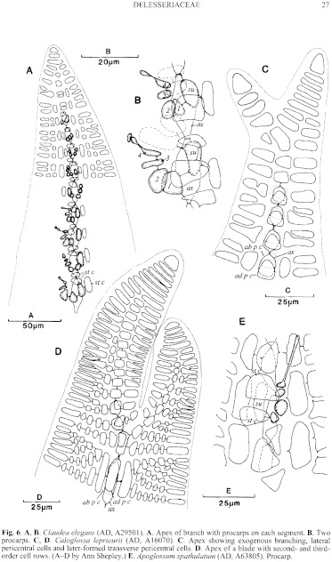
Figure 6 enlarge
Fig. 6. A, B. Claudea elegans (AD, A29501). A. Apex of branch with procarps on each segment. B. Two procarps. C, D. Caloglossa leprieurii (AD, A16070). C. Apex showing exogenous branching, lateral pericentral cells and later-formed transverse pericentral cells. D. Apex of a blade with second- and third-order cell rows. (A–D by Ann Shepley.) E. Apoglossum spathulatum (AD, A63805). Procarp.
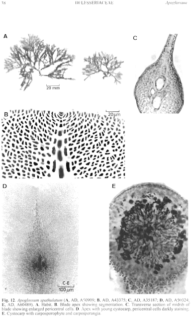
Figure 12 enlarge
Fig. 12. Apoglossum spathulatum (A, AD, A50909; B, AD, A43375; C, AD, A35187; D, AD, A50324; E, AD, A60489). A. Habit. B. Blade apex showing segmentation. C. Transverse section of midrib of blade showing enlarged pericentral cells. D. Apex with young cystocarp, pericentral cells darkly staining. E. Cystocarp with carposporophyte and carposporangia.
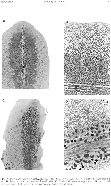
Figure 13 enlarge
Fig. 13. Apoglossum spathulatum (A, B, AD, A43375; C, D, AD, A60489). A. Blade with spermatangial sori. B. Spermatangial sori between lateral veins. C. Blade with tetrasporangial sorus. D. Sorus with tetrasporangia cut off from inner cells of second- and third-order rows.

|
Email Contact: State Herbarium of South Australia |

|


