|
|
|
|
|
|||||||||||
|
Electronic Flora of South Australia Species Fact Sheet
Phylum Rhodophyta – Order Ceramiales – Family Ceramiaceae – Tribe Pterothamnieae
Selected citations: Athanasiadis 1996: 64, fig. 17.
Synonym
Platythamnion cuspidatum Wollaston 1978: 3, figs 1–9, 15–17. Millar & Kraft 1993: 41.
Thallus (Fig. 60E) red, erect, 1–2 cm high, complanately branched with lateral branches 7–10 axial cells apart, upper axes flexuous (Fig. 60E), ecorticate throughout, basally attached by rhizoids from axial cells; epizoic or epiphytic. Structure. Apical cells 6–11 µm in diameter and L/D 1–1.6, enlarging to 60–140 µm in diameter and L/D 1–2 in lower axes. Axial cells each with 4 whorl-branchlets, 2 opposite major ones (Fig. 60E) 200–250 µm long in the plane of the thallus, and 2 smaller alternating minor ones 50–130 µm long; major pinnae with simple or branched pinnules directed upwards, tapering to an acute terminal cell, basal cells of rachides 20–35 µm in diameter and L/D 1–1.5, tapering to subterminal cells 4–8 µm in diameter and L/D 1–2, apical cells mucronate; minor pinnae simple or branched, often with 3–4 slender, spinous, branches (Fig. 61L); gland cells (Fig. 60E) lateral on mid cells of rachides or pinnules, ovoid. 12–18 µm in diameter. Lateral branches arising in place of a major pinna. Cells (smaller) uninucleate, rhodoplasts discoid in small cells, becoming ribbon like in larger cells.
Reproduction: Gametophytes dioecious. Procarps not observed. Carposporophytes terminal. Spermatangia terminal in branched clusters (Figs 60F, 61M) on cells of pinnules.
Tetrasporangia (Figs 60G, 61N) occur on cells of pinnules or their branches, sessile or terminal, subspherical, 20–30 µm in diameter, decussately divided.
Type from Port Kembla, N.S.W., 20 m deep (Watson, 16.ix.1976); holotype in AD, A47994.
Selected specimens: Portland, Vic., 7–9 m deep (Kraft 8266a, 27.iv.1990; MELU and AD, A61243). Gabo I., Vic., on Carpoglossum, 28 m deep (Shepherd, 19.ii.1973; AD, A43550).
Distribution: Port Kembla, N.S.W., and Portland and Gabo I., Victoria.
References:
ATHANASIADIS, A. & KRAFT, G.T. (1994). Description of Pterothamnion squarrulosum (Harvey) comb. nov. from south-eastern Australia and southern New Zealand, with a taxonomic re-assessment of the genera Pterothamnion, Platythamnion & Glandothamnus (Ceramiaceae, Rhodophyta). Eur. J. Phycol. 29, 119–133.
ATHANASIADIS, A. (1996). Morphology and classification of the Ceramioideae (Rhodophyta) based on phylogenetic principles. Opera Botanica No. 128, pp. 1–216.
MILLAR, A.J.K. & KRAFT, G.T. (1993). Catalogue of marine and freshwater Red Algae (Rhodophyta) of New South Wales, including Lord Howe Island, South-western Pacific. Aust. Syst. Bot. 6, 1–90.
WOLLASTON, E.M. (1978). Two new species of Platythamnion J. Agardh (Ceramiaceae, Rhodophyta) from eastern and southern Australia. Trans. R. Soc. S. Aust. 102, 1–8.
The Marine Benthic Flora of Southern Australia Part IIIC complete list of references.
Publication:
Womersley, H.B.S. (24 December, 1998)
The Marine Benthic Flora of Southern Australia
Rhodophyta. Part IIIC. Ceramiales – Ceramiaceae, Dasyaceae
©State Herbarium of South Australia, Government of South Australia
Illustrations in Womersley Part IIIA, 1998: FIGS 60 E–G, 61 L–N.
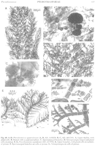
Figure 60 enlarge
Fig. 60. A–D. Pterothamnion squarrulosum (A, D, AD, A48820; B, C, AD, A61244). A. Upper thallus, with gland cells. B. Branch with a carposporophyte. C. Spermatangial branches. D. Tetrasporangia (larger) and gland cells on pinnae. E–G.Pterothamnion cuspidatum (AD, A47994). E. Upper branches, with gland cells on rachides of pinnae. F. Spermatangial branches on cells of pinnae. G. Tetrasporangia on cells of pinnae.
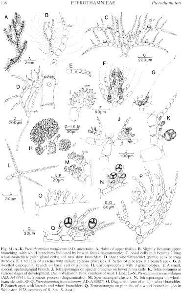
Figure 61 enlarge
Fig. 61. A–K. Pterothamnion nodiferum (AD, uncertain). A. Habit of upper thallus. B. Slightly flexuous upper branching, with whorl-branchlets indicated by broken lines (diagrammatic). C. Axial cells each bearing 2 long whorl-branchlets (with gland cells) and two short branchlets. D. Inner whorl-branchlet (pinna) cells bearing rhizoids. E. End cells of a rachis with minute spinous processes. F. Series of procarps at a branch apex. G. A 4-celled carpogonial branch on basal cell of a pinna. H. Carposporophyte with 3 gonimolobes. I. A small, special, spermatangial branch. J. Tetrasporangia on special branches on lower pinna cells. K. Tetrasporangia at various stages of development. (As in Wollaston 1968, courtesy of Aust. J. Bot.) L–N. Pterothamnion cuspidatum (AD, A47994). L. Spinous process (diagrammatic). M. Spermatangial clusters. N. Tetrasporangia on whorl-branchlet cells. O–Q. Pterothamnion francisianum (AD, A38087). O. Diagram of form of a major whorl-branchlet. P. Branch apex with laterals and whorl-branchlets. Q. Tetrasporangia on pinnules of a whorl-branchlet. (As in Wollaston 1978, courtesy of R. Soc. S. Aust.)

|
Email Contact: State Herbarium of South Australia |

|


