|
|
|
|
|
|||||||||||
|
Electronic Flora of South Australia Species Fact Sheet
Phylum Rhodophyta – Order Ceramiales – Family Ceramiaceae – Tribe Rhodocallideae
Selected citations: Huisman & Walker 1990: 424. Lucas 1909: 51. Lucas & Perrin 1947: 346. May 1965: 375. Silva et al. 1996: 421.
Synonyms
Ptilota siliculosa Harvey 1855a: 559; 1863, synop.: xlix. J. Agardh 1876: 79. Sonder 1881: 11.
Plumaria siliculosa (Harvey) Kuntze 1891: 911.
Thallus (Fig. 170A) erect, dark red-brown, 4–10 cm high, pinnate, much branched complanately with compressed indeterminate branches bearing alternate, close set, slightly compressed, tapering determinate branchlets 1–2 mm long, branches 0.6–1.2 mm broad, not striate. Holdfast small, rhizoidal; epiphytic, usually on Nizymenia conferta (Stenocladia australis). Structure. Apical cells (Fig. 170C) 10–20 µm in diameter and L/D about 1, cutting off periaxial cells from subapical cells (Fig. 170B, C) and becoming enclosed by developing determinate branchlets, axial cells increasing to 90–120 µm in diameter and L/D 1.5–2.5, with lateral filaments of determinate branchlets lying in the plane of branching, alternate every (2–) 3 (–4) axial cells, with 2 transverse periaxial cells (Fig. 170D) on each side of the branch; periaxial filaments not extending beyond the compact cortex. Central part of older branches becoming swollen by slight to considerable rhizoid development around the axial cells (Fig. 170D); surface cells of the cortex small, compact, isodiametric, 2–3 µm across. Cells uninucleate; rhodoplasts discoid, in chains in larger cells.
Reproduction: Carpogonial branches unknown. Carposporophytes (Fig. 170E) formed in dense globular tufts 300–600 µm across, with a short multicellular base and curved surface filaments 90–180 µm long and 10–12 µm in diameter, cells L/D 0.8–1.5; gonimolobes several in each tuft, rounded, 120–180 µm across, with ovoid carposporangia 8–14 µm in diameter. Spermatangia unknown.
Tetrasporangia produced in siliculose branchlets (Fig. 170F) adaxial on determinate branchlets, each branchlet with an axial row of 12–20 cells bearing lateral cell rows and a surface layer of short 8–14-celled filaments, among which occur the slightly ovoid terminal (on 1–3-celled pedicels) tetrasporangia, 25–28 µm in diameter, tetrahedrally divided.
Type from Rottnest I., W. Aust.(Harvey); lectotype Tray. Set 243, Herb. Harvey, TCD.
Selected specimens: Champion Bay, W. Aust. (Algae Muellerianae; MEL 26397). Flat Rocks, 40 km S of Geraldton, W. Aust., on *Nizymenia conferta, drift (Mitchell, 17.ix.1966; AD, A31043). Point Peron, W. Aust., drift (Royce 966, 21.iv.1951; AD, A16216). Israelite Bay, W. Aust. ("Algae Muellerianae", MEL, 26398). Vivonne Bay, Kangaroo I., S. Aust., drift (Womersley, 2.i.1949; AD, A10611). Seal Bay, Kangaroo I., S. Aust., on Nizymenia conferta, drift (Womersley, 22.xi.1968; AD, A32960). Robe, S. Aust., drift (Womersley, 15.iv.1959; AD, A23030). Warrnambool, Vic., drift (Womersley, 13.iv.1959; AD, A22427). Point Roadknight, Vic., reef pools (Womersley, 6.vi.1953; AD, A18765).
Distribution: Champion Bay, W. Aust., to Point Roadknight, Victoria.
References:
AGARDH, J.G. (1876). Species Genera et Ordines Algarum. Vol. 3, Part 1- Epicrisis systematic Floridearum, pp. i-vii, 1–724. (Weigel: Leipzig.)
DE TONI, G.B. (1903). Sylloge Algarum omnium hucusque Cognitarum. Vol. 4. Florideae. Sect. 3, pp. 775–1521 + 1523–1525. (Padua.)
HARVEY, W.H. (1855a). Some account of the marine botany of the colony of Western Australia. Trans. R. Jr. Acad. 22, 525–566.
HARVEY, W.H. (1863). Phycologia Australica. Vol. 5, Plates 241–300, synop., pp. i-lxxiii. (Reeve: London.)
HUISMAN, J.M. & WALKER, D.I. (1990). A catalogue of the marine plants of Rottnest Island, Western Australia, with notes on their distribution and biogeography. Kingia 1, 349–459.
KUNTZE, O. (1891). Revisio generum Plantarum. Part II. 4. Algae, pp. 877–930. (Leipzig.)
LUCAS, A.H.S. & PERRIN, F. (1947). The Seaweeds of South Australia. Part 2. The Red Seaweeds. (Govt Printer: Adelaide.)
LUCAS, A.H.S. (1909). Revised list of the Fucoideae and Florideae of Australia. Proc. Linn. Soc. N.S.W. 34, 9–60.
MAY, V. (1965). A census and key to the species of Rhodophyceae (red algae) recorded from Australia. Contr. N.S.W. natn. Herb. 3, 349–429.
SILVA, P.C., BASSON, P.W. & MOE, R.L. (1996). Catalogue of the Benthic Marine Algae of the Indian Ocean. (University of California Press: Berkeley, Los Angeles & London.)
SONDER, O.W. (1881). In Mueller, F., Fragmenta Phytographiae Australiae. Supplementum ad volumen undecinum: Algae Australianae hactenus cognitae, pp. 1–42, 105–107. (Melbourne.)
The Marine Benthic Flora of Southern Australia Part IIIC complete list of references.
Publication:
Womersley, H.B.S. (24 December, 1998)
The Marine Benthic Flora of Southern Australia
Rhodophyta. Part IIIC. Ceramiales – Ceramiaceae, Dasyaceae
©State Herbarium of South Australia, Government of South Australia
Illustration in Womersley Part IIIA, 1998: FIG. 170.
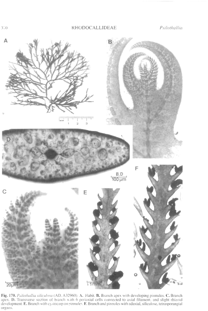
Figure 170 enlarge
Fig. 170. Psilothallia siliculosa (AD, A32960). A. Habit. B. Branch apex with developing pinnules. C. Branch apex. D. Transverse section of branch with 6 periaxial cells connected to axial filament, and slight rhizoid development. E. Branch with cystocarp on pinnules. F. Branch and pinnules with adaxial, siliculose, tetrasporangial organs.

|
Email Contact: State Herbarium of South Australia |

|


