|
|
|
|
|
|||||||||||
|
Electronic Flora of South Australia Species Fact Sheet
Phylum Rhodophyta – Order Ceramiales – Family Ceramiaceae – Tribe Ceramieae
Selected citations: Huisman 1997: 197. Huisman et al. 1990: 96. Kendrick et al. 1990: 51. Millar & Kraft 1993: 39. Silva et al. 1996: 403.
Thallus (Fig. 176A–C) grey-red to red, 2–12 mm high, erect filaments sparsely subdichotomous or lateral, basal filaments prostrate, attached by rhizoids from periaxial cells or their derivatives, 1–3 cells long and with multicellular pads; epiphytic on Amphibolis, Posidonia or on algae associated with these seagrasses. Structure. Branches 70–150 µm in diameter, tapering only slightly above, with relatively straight apices (Fig. 176B); axial cells usually L/D 0.7–2, with narrow nodal bands 2 cells long and internodal spaces 3–6 times as long as nodal bands (Fig. 176A–C). Periaxial cells usually 6 (Fig. 177A, B), each cutting off laterally one (occasionally 2, rarely none) pseudoperiaxial cell which lies in the periaxial ring (Fig. 177B), and each cell of this ring then cutting off acropetally a single cortical cell, thus forming a nodal band 2 cells long (Fig. 177A). Each direct derivative of the true periaxial cells producing a short tapering filament (55–100 µm long) of 3–6 (–7) cells (with a rounded apex) which projects outwardly and forward from the axis (Figs 176A–C, 177A, B); the true periaxial cells usually do not produce such a filament, though both they and the acropetal derivatives of the pseudoperiaxial cells may do so; slender terminal hairs often present on these filaments. Rhodoplasts discoid in cortical cells, linear in axial cells.
Reproduction: Gametophytes dioecious. Carposporophytes (Fig. 176A) globular, 120–180 (–200) µm across, with angular to ovoid carposporangia 30–40 µm across, subtended by 1–3 short branchlets. Spermatangial masses (Fig. 176B) cut off first from adaxial nodal cells, later developing all around the nodes.
Type from 4 km S of Redcliff Point, N Spencer Gulf, S.Aust., on Posidonia sinuosa, 10 m deep (Johnson, 10.i.1977); holotype in AD, A47850.
Selected specimens: Port Denison, W. Aust., on Erythroclonium sedoides, drift (Kraft, 14.xii.1971; AD, A41279). Coffin Bay, S. Aust., on Posidonia australis, 2.5 m deep (Womersley, 4.xii.1975; AD, A46952). Redcliff Point, N Spencer Gulf, S. Aust., on Posidonia australis, 7 m deep (Johnson, 17.iv.1975: AD, A47825). Tiparra Reef, S. Aust., on Amphibolis antarctica, 11m deep (Shepherd, 5.xi.1971; AD, A38322). Sellicks Beach, S. Aust., on Haliptilon roseum, drift (O'Leary, 12.vi.1993; AD, A62187). 3 km SW of Kingston, S. Aust., on Amphibolis antarctica, 6–7m deep (R. Lewis, 28.xi.1972; AD, A42881). Snowy River Mouth, Vic. (Mueller, Feb 1855; MEL, 45455).
Distribution: Shark Bay, W. Aust., (Kendrick et al. 1990, p. 51), to Port Stephens, N.S.W. and Lord Howe I. (Millar & Kraft 1993, p. 39).
Taxonomic notes: Tetrasporangia (Figs 176C, 177A, B) cut off from enlarged periaxial cells, usually single per node and abaxial, often with further sporangia formed later from adjacent cells, protected by slightly greater development of nodal filaments (some basally dichotomous) than in sterile plants; tetrasporangia 50–70 µm in diameter, tetrahedrally to decussately divided.
All collections (except MEL 45455, probably drift) have been associated with seagrass beds, between 2.5 and 12m deep, in areas of moderate water movement. At Redcliff Point it occurs 2–10m deep on Posidonia, throughout the year but most commonly in summer and autumn (December-June) and least in spring.
C. shepherdii is well marked by the irregular double whorls of filaments at the nodes and the pattern of nodal development. In forming pseudoperiaxial cells it shows similarity to C. australe, which is otherwise quite distinct. The blunt apices of the filaments separate C. shepherdii from C. monacanthum and C. puberulum and from the numerous extra-Australian species with acute spines, none of which are similar in their nodal development.
References:
HUISMAN, J.M. (1997). Marine Benthic Algae of the Houtman Abrolhos Islands, Western Australia. In Wells, F.E. (Ed.) The Marine Flora and Fauna of the Houtman Abrolhos Islands, Western Australia, pp. 177–237. (W. Aust. Museum: Perth.)
HUISMAN, J.M., KENDRICK, G.A., WALKER, D.I. & COUTÉ, A. (1990). The Marine Algae of Shark Bay, Western Australia. Research in Shark Bay. Report of the France-Australe Bicentenary Expedition Committee, pp. 89–100.
KENDRICK, G.A., HUISMAN, J.M. & WALKER, D.I. (1990). Benthic macroalgae of Shark Bay, Western Australia. Bot. Mar 33, 47–54.
MILLAR, A.J.K. & KRAFT, G.T. (1993). Catalogue of marine and freshwater Red Algae (Rhodophyta) of New South Wales, including Lord Howe Island, South-western Pacific. Aust. Syst. Bot. 6, 1–90.
SILVA, P.C., BASSON, P.W. & MOE, R.L. (1996). Catalogue of the Benthic Marine Algae of the Indian Ocean. (University of California Press: Berkeley, Los Angeles & London.)
WOMERSLEY, H.B.S. (1978). Southern Australian species of Ceramium Roth (Rhodophyta). Aust. J. Mar. Freshw. Res. 29, 205–257.
The Marine Benthic Flora of Southern Australia Part IIIC complete list of references.
Publication:
Womersley, H.B.S. (24 December, 1998)
The Marine Benthic Flora of Southern Australia
Rhodophyta. Part IIIC. Ceramiales – Ceramiaceae, Dasyaceae
©State Herbarium of South Australia, Government of South Australia
Illustrations in Womersley Part IIIA, 1998: FIGS 176 A–C, 177A, B.
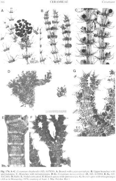
Figure 176 enlarge
Fig. 176. A–C. Ceramium shepherdii (AD, A47850). A. Branch with carposporophyte. B. Upper branches with spermatangia. C. Branches with tetrasporangia. D–G. Ceramium monacanthum (D, AD, A32694; E–G, AD, A42260). D. Habit. E. Nodal cortication. F. Branch apices with spermatangia. G. Branch apex with tetrasporangia (All as in Womersley 1978, courtesy of Aust. J. Mar. Freshw. Res.)
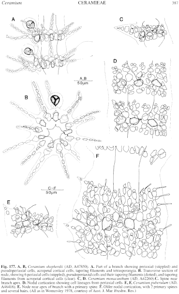
Figure 177 enlarge
Fig. 177. A, B, Ceramium shepherdii (AD, A47850). A. Part of a branch showing periaxial (stippled) and pseudoperiaxial cells, acropetal cortical cells, tapering filaments and tetrasporangia. B. Transverse section of node, showing 6 periaxial cells (stippled), pseudoperiaxial cells and their tapering filaments (dotted), and tapering filaments from acropetal cortical cells (clear). C, D. Ceramium mortacanthum (AD, A42260).C. Spine near branch apex. D. Nodal cortication showing cell lineages from periaxial cells. E, F, Ceramium puberulum (AD, A46416). E. Node near apex of branch with a primary spine. F. Older nodal cortication, with 2 primary spines and several hairs. (All as in Womersley 1978, courtesy of Aust. J. Mar. Freshw. Res.)

|
Email Contact: State Herbarium of South Australia |

|


