|
|
|
|
|
|||||||||||
|
Electronic Flora of South Australia Species Fact Sheet
Phylum Rhodophyta – Class Florideophyceae – Order Corallinales – Family Corallinaceae – Subfamily Melobesioideae
Synonyms
Lithothamnion repandum Foslie 1904b: 4. Woelkerling 1993: 189.
Leptophytum repandum (Foslie) Adey 1970: 30.
Lithothamnion lenormandii f. australis Foslie 1901a: 8. Woelkerling 1993: 36.
Lithothamnion absonum Foslie 1907b: 6–7. Woelkerling 1993: 14.
Leptophytum absonum (Foslie) Adey 1970: 29.
Lithothamnion repandum f. asperula Foslie 1906: 5. Woelkerling 1993: 31.
Lithothamnion asperulum (Foslie) Foslie 1907b: 6.
Leptophytum asperulum (Foslie) Adey 1970: 29.
Additional references involving these binomials and information on synonymy are provided by Wilks & Woelkerling 1994.
Thallus normally dull pinkish, encrusting to warty to fruticose, mostly 4–80 mm across and 0.1–4.0 mm thick or tall, epigenous and completely affixed by cell adhesion; protuberant branches, simple or branched, mostly 1–6 mm in diameter and 1–4 mm long. Structure pseudoparenchymatous; organisation dorsiventral in crustose portions but radial in protuberant branches; construction monomerous, consisting of a single system of branched, laterally cohering, filaments that collectively contribute to a ventrally situated core in crustose portions or centrally situated core in protuberant branches and a peripheral region where portions of core filaments or their derivatives curve outwards towards the thallus surface, each filament composed of cells 2–12 µm in diameter and 2–27 µm long; epithallial cells 2–8 µm in diameter and 1–8 µm long, terminating most filaments at the thallus surface, with distal walls rounded or flattened but not flared; cell elongation occurring mainly behind actively dividing subepithallial initials that are usually as short as or shorter than their immediate inward derivatives; cells of adjacent filaments joined by cell-fusions; secondary pit-connections, haustoria, and trichocytes unknown.
Reproduction: Vegetative reproduction unknown. Gametangia and carposporophytes produced in uniporate conceptacles; tetrasporangia and bisporangia produced in multiporate conceptacles.
Gametangial thalli monoecious or dioecious; carpogonia and spermatangia produced in separate conceptacles or occasionally in the same conceptacle. Carpogonia terminating 2- or 3-celled filaments arising from the female conceptacle chamber floor. Mature female carposporangial conceptacle roofs protruding above or flush with surrounding thallus surface, 22–100 µm thick, composed of 5–10 layers of cells above the chamber, conceptacle chambers 112–300 µm in diameter and 50–125 µm high. Mature carposporophytes apparently lacking a conspicuous central fusion cell and consisting of several-celled gonimoblast filaments bearing terminal carposporangia 12–75 µm in diameter. Both unbranched and branched spermatangial filaments present, arising from the floor, walls and roof of male conceptacle chambers, mature male conceptacle roofs protruding above surrounding thallus surface, 25–75 µm thick, composed of 4–9 layers of cells above the chamber, conceptacle chambers 100–250 µm in diameter and 45–150 µm high.
Tetrasporangial/bisporangial conceptacle roofs protruding above surrounding surface, 3–5 cells thick above the chamber, pore canals lined by cells that are similar in size and shape to other roof cells; conceptacle chambers 96–300 µm in diameter and 50–150 µm high, not filled with enlarged irregularly shaped vegetative cells interspersed amongst the sporangia and lacking distinct layers of darkly staining cells beneath the chamber floor; tetrasporangia scattered across the conceptacle chamber floor, each mature sporangium 15–80 µm in diameter and 32–125 µm long, containing zonately arranged tetraspores and possessing an apical plug that blocks a roof pore prior to spore release; bisporangia occasional, 15–80 µm in diameter and 32–125 µm long.
Type from Half Moon Bay, Port Phillip Bay, Vic. (Gabriel, 14.i.1899); lectotype in TRH (unnumbered; includes slides 358 and 516); designated by Adey in Adey & Lebednik (1967, p. 83); depicted in Wilks & Woelkerling (1994, p. 206, fig. 1); additional data provided by Woelkerling (1993, p. 189).
Selected specimens: Reef 10.4 km E of Eyre, W. Aust., 0–2 m deep (Woelkerling, Platt & Jones, 3.ii.1984; LTB, 14122, 14123, 14141). Point Sinclair, S. Aust., 0–2 m deep (Woelkerling, Platt & Jones, 15.ii.1984; LTB, 14521). Hansen Bay, Kangaroo I., S. Aust., 0–1.5 m deep (Campbell & Penrose, 9.iv.1988; LTB, 15704, 15708). Beachport (reef W of Schnapper Point), S. Aust., 0–2 m deep (Campbell, Penrose & Woelkerling, 26.ii.1988; LTB, 15788, 15795). Beachport (Post Office Rock), S. Aust., 0–3 m deep (Campbell & Penrose, 6.xi.1987; LTB, 15836). Beachport (Woolley's Rocks), S. Aust., intertidal (Penrose & Woelkerling, 25.ii.1991; LTB, 16204, 16205, 16207, 16208). Blanket Bay, Otway National Park, Vic., 4 m deep (Campbell, Penrose & May, 14.xi.1985; LTB, 15235). Rye (Number 16 reef) (Woelkerling & Wilks, Feb. 1991; LTB, 16209). Flinders, Western Port, Vic., reef pool (Woelkerling, 13.i.1983; LTB, 12650). Kitty Miller Bay, Phillip I., Vic., intertidal (Woelkerling, Penrose & Wilks, 12.iv.1991; LTB, 16167, 16175, 16180, 16181). Cape Conran, Vic., reef pool (Platt, 29.xii.1982; LTB, 12633). Greens Beach, Tas., intertidal pool (Platt, 2.iii.1983; LTB, 13414). Binalong Bay, Bay of Fires, Tas., 0–2 m deep (Platt & Woelkerling, 23.ii.1983; LTB, 13203, 13194). Tessellated Pavement, Eaglehawk Neck, Tas., 1–2 m deep (Platt & Woelkerling, 26.ii.1983; LTB, 13247, 13250).
Distribution: In Australia, from 10 km east of Eyre, W. Aust., to Cape Conran, Vic., and around Tasmania.
New Zealand.
Taxonomic notes: Phymatolithon repandum occurs intertidally on reef surfaces and in pools and is known subtidally to depths of 6 m in southern Australia. Thalli have been found on rock and glass; epiphytic and epizoic thalli have not been encountered. Thalli with young tetrasporangial and bisporangial conceptacles can sometimes be recognised in the field because senescent vegetative discs above developing conceptacles (Fig. 80E) lose pigmentation, turn white, and form conspicuous dots on the thallus surface. Tentative field identifications made on this basis need to be confirmed by sectioning.
References:
ADEY, W.H. & LEBEDNIK, P.A. (1967). Catalog of the Foslie Herbarium. (Det Kongelige Norske Videnskabers Selskab Museet: Trondheim, Norway.)
ADEY, W.H. (1970). A revision of the Foslie crustose coralline herbarium. K. norske Vidensk. Selsk. Skr. 1970 (1), 1–46.
FOSLIE, M. (1901a). New melobesieae. K. norske Vidensk. Selsk. Skr. 1900(6), 1–24.
FOSLIE, M. (1904b). Algologiske notiser. K. norske Vidensk. Selsk. Skr. 1904(2), 1–9.
FOSLIE, M. (1906). Algologiske notiser II. K. norske Vidensk. Selsk. Skr. 1906(2), 1–28.
FOSLIE, M. (1907b). Algologiske notiser. IV. K. norske Vidensk. Selsk. Skr. 1907(6), 1–30.
WILKS, K.M. & WOELKERLING, W.J. (1994). An account of southern Australian species of Phymatolithon (Corallinaceae, Rhodophyta) with comments on Leptophytum. Aust. Syst. Bot. 7, 183–223.
WOELKERLING, Wm.J. (1993). Type collections of Corallinales (Rhodophyta) in the Foslie Herbarium (TRH). Gunneria 67, 1–289.
The Marine Benthic Flora of Southern Australia Part IIIB complete list of references.
Publication:
Womersley, H.B.S. (28 June, 1996)
The Marine Benthic Flora of Southern Australia
Rhodophyta. Part IIIB. Gracilarialse, Rhodymeniales, Corallinales and Bonnemaisoniales
Reproduced with permission from The Marine Benthic Flora of Southern Australia Part IIIB 1996, by H.B.S. Womersley. Australian Biological Resources Study, Canberra. Copyright Commonwealth of Australia.
Illustrations in Womersley Part IIIA, 1996: PLATE 3 fig. 4; FIGS 80, 81.
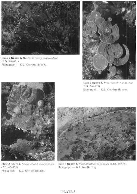
Plate 3 enlarge
PLATE 3
figure 1. Mastophoropsis canaliculata (AD, 64421). Photograph K.L. Gowlett-Holmes.
figure 2. Phymatolithon masonianum (AD, A64476). Photograph - K.L.Gowlett-Holmes.
figure 3. Synarthrophyton patena (AD, A64409). Photograph - K.L.Gowlett-Holmes.
figure 4. Phymatolithon repandum (LTB, 15836). Photograph - W.J. Woelkerling.
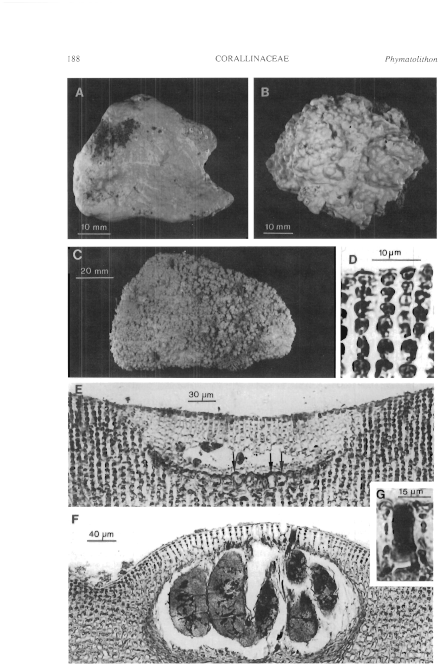
Figure 80 enlarge
Fig. 80. Phymatolithon repandum (A, LTB, 16209; B, F, LTB, 15836; C, LTB, 15795; D, E, G, LTB, 16205). A. Encrusting thallus on rock. B. Warty thallus on rock. C. Fruticose thallus on rock. D. Section of thallus showing epithallial cells and subepithallial initials that are as short as or shorter than the cells subtending them. E. Section of very young tetrasporangial conceptacle arising adventitiously from a group of vegetative cells (arrows) within the thallus. F. Section of mature tetrasporangial conceptacle. G. Portion of tetrasporangial conceptacle roof showing pore canals and bordering filaments.
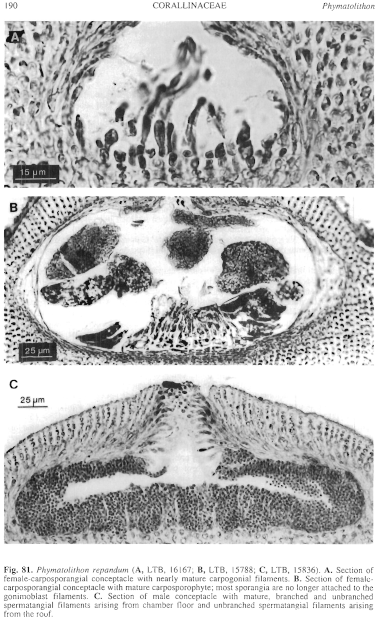
Figure 81 enlarge
Fig. 81. Phymatolithon repandum (A, LTB, 16167; B, LTB, 15788; C, LTB, 15836). A. Section of female-carposporangial conceptacle with nearly mature carpogonial filaments. B. Section of female-carposporangial conceptacle with mature carposporophyte; most sporangia are no longer attached to the gonimoblast filaments. C. Section of male conceptacle with mature, branched and unbranched spermatangial filaments arising from chamber floor and unbranched spermatangial filaments arising from the roof.

|
Email Contact: State Herbarium of South Australia |

|


