|
|
|
|
|
|||||||||||
|
Electronic Flora of South Australia Species Fact Sheet
Phylum Rhodophyta – Class Florideophyceae – Order Corallinales – Family Corallinaceae – Subfamily Melobesioideae
Thallus normally pinkish, encrusting to warty to fruticose, mostly 40–95 mm across and 0.2–25 mm thick or tall, epigenous and partially to completely affixed by cell adhesion; protuberant branches simple or branched, mostly 1.4–11 mm in diameter and 1.8–14 mm long. Structure pseudoparenchymatous, organisation dorsiventral in crustose portions but radial in protuberant branches; construction monomerous, consisting of a single system of branched, laterally cohering, filaments that collectively contribute to a ventrally or centrally situated core and a peripheral region where portions of core filaments or their derivatives curve outwards towards the thallus surface, each filament composed of cells 4–12 µm in diameter and 6–42 µm long; epithallial cells 3–5 µm in diameter and 4–6 µm long, terminating most filaments at the thallus surface, with distal walls rounded or flattened but not flared; cell elongation occurring mainly within actively dividing subepithallial initials that are usually as long as or longer than their immediate inward derivatives; cells of adjacent filaments joined by cell-fusions; trichocytes sometimes present, solitary or in small fields; secondary pit-connections and haustoria unknown.
Reproduction: Vegetative reproduction unknown. Gametangia and presumably carposporophytes produced in uniporate conceptacles; tetrasporangia produced in multiporate conceptacles. Bisporangia unknown.
Gametangial thalli probably dioecious; carpogonia and spermatangia produced in separate conceptacles. Carpogonial stages and carposporophytes unknown. Spermatangial filaments unbranched, arising from the floor, walls and roof of male conceptacle chambers; mature male conceptacle roofs protruding above surrounding thallus surface, 54–94 µm thick, composed of 11–13 layers of cells above the chamber, conceptacle chambers 175–243 µm in diameter and 48–60 µm high.
Tetrasporangial conceptacle roofs protruding above surrounding surface, differentiated into a peripheral rim and a central sunken pore-plate, 3–6 cells thick above the chamber, pore canals lined by narrow elongate cells that differ in shape from other roof cells especially near the base of the canal, conceptacle chambers 185–420 µm in diameter and 175–190 µm high; tetrasporangia scattered across the conceptacle chamber floor; each mature sporangium 54–122 µm in diameter and 121–150 µm long, containing zonately arranged tetraspores and possessing an apical plug that blocks a roof pore prior to spore release.
Type from Blanket Bay, Otway National Park, Vic. (Campbell, Penrose & May, 14.xi.1985); holotype in LTB (15249); depicted in Woelkerling & Harvey (1993, p. 630, fig. 24A).
Selected specimens: Beachport (Post Office Rock), S. Aust., on reef, 0–3 m deep (Campbell, Penrose & Woelkerling, 1.xii.1986; LTB, 15507; Campbell & Penrose, 6.xi.1987; LTB, 15834 and Harvey & Condon, 15.ii.1993; LTB, 16594). Beachport (platform west of Post Office Rock), S. Aust., 0–1 m deep (Woelkerling, 6.xi.1987; LTB, 15821). Blanket Bay, Otway National Park, Vic., 2 m deep (Campbell, Penrose & May, 14.xi.1985; LTB, 15257). Rye (Number Sixteen reef), Vic., reef pool (Platt & Woelkerling, 21.xii.1982; LTB, 12622). Summerland Bay, Phillip I., Vic., 1–2 m deep (Harvey & Harvey, 22.ii.1993; LTB, 16591, 16592). Binalong Bay, Tas., 0–4 m deep (Plan, 23.ii.1983; LTB, 13207, 13212). Eaglehawk Neck (Clydes I.), Tas., 0–6 m deep (Platt & Woelkerling, 26.ii.1983; LTB, 13297). Safety Cove, Port Arthur, Tas., 0–2 m deep (Platt, 25.ii.1983; LTB, 12859 and Plan & Woelkerling, 25.ii.1983; LTB, 12850). Variety Bay (S shore), Bruny I., Tas., 2–4 m deep (Platt, 17.ii.1983;LTB, 13008).
Distribution: Beachport, S. Aust., to Summerland Bay, Phillip I., Vic., and the eastern and southern coasts of Tasmania.
Taxonomic notes: Thalli of Mesophyllum printzianum have been found in intertidal rock pools and to depths of 6 m on rock and on the holdfasts of the brown alga Phyllospora.
References:
WOELKERLING, W.J. & HARVEY, A. (1993). An account of sourthern Australian species of Mesophyllum (Corallinaceae, Rhodophyta). Aust. Syst. Bot. 6, 571–637.
The Marine Benthic Flora of Southern Australia Part IIIB complete list of references.
Publication:
Womersley, H.B.S. (28 June, 1996)
The Marine Benthic Flora of Southern Australia
Rhodophyta. Part IIIB. Gracilarialse, Rhodymeniales, Corallinales and Bonnemaisoniales
Reproduced with permission from The Marine Benthic Flora of Southern Australia Part IIIB 1996, by H.B.S. Womersley. Australian Biological Resources Study, Canberra. Copyright Commonwealth of Australia.
Illustrations in Womersley Part IIIA, 1996: FIGS 63B, 64C, 88, 89.
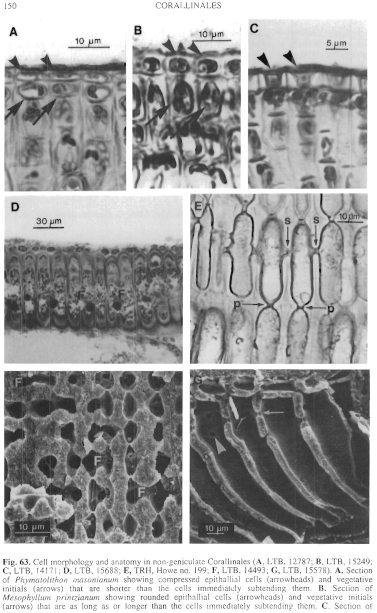
Figure 63 enlarge
Fig. 63. Cell morphology and anatomy in non-geniculate Corallinales (A, LTB, 12787; B, LTB, 15249; C, LTB, 14171; D, LTB, 15688; E, TRH, Howe no. 199; F, LTB, 14493; G, LTB, 15578). A. Section of Phymatolithon masonianum showing compressed epithallial cells (arrowheads) and vegetative initials (arrows) that are shorter than the cells immediately subtending them. B. Section of Mesophyllum printzianum showing rounded epithallial cells (arrowheads) and vegetative initials (arrows) that are as long as or longer than the cells immediately subtending them. C. Section of Sporolithon durum showing epithallial cells (arrow heads) with flared outer walls D. Palisade cells of contiguous filaments of Metamastophora flabellata showing cell-fusions (F). E. Columnar cells of Lithophyllum frondosum showing primary (arrow - p) and secondary (arrow - s) pit connections. F. Fracture of Sporolithon durum showing fusions (F) between cells of contiguous filaments. G. Fracture of Lithophyllum corallinae showing pit connections in surface (arrowhead) and sectional (arrow) views.
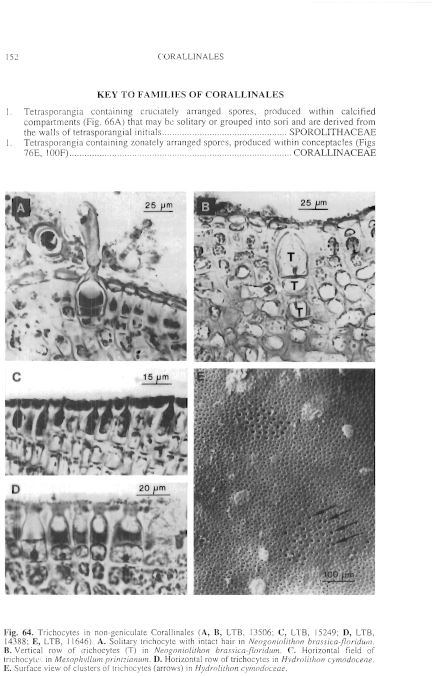
Figure 64 enlarge
Fig. 64. Trichocytes in non-geniculate Corallinales (A, B, LTB, 13506; C, LTB, 15249; D, LTB, 14388; E, LTB, 11646). A. Solitary trichocyte with intact hair in Neogoniolithon brassica-floridum. B. Vertical row of trichocytes (T) in Neogoniolithon brassica-floridum. C. Horizontal field of trichocytes in Mesophyllum printzianum. D. Horizontal row of trichocytes in Hydrolithon cymodoceae. E. Surface view of clusters of trichocytes (arrows) in Hydrolithon cymodoceae.
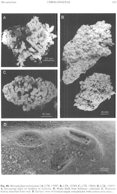
Figure 88 enlarge
Fig. 88. Mesophyllum printzianum (A, LTB, 13297; B, LTB, 15249; C, LTB, 15834; D, LTB, 15507). A. Encrusting thalli on holdfast of Ecklonia. B. Warty thalli from holotype collection. C. Fruticose thallus detached from rock. D. Surface view of tetrasporangial conceptacles with sunken pore-plates.
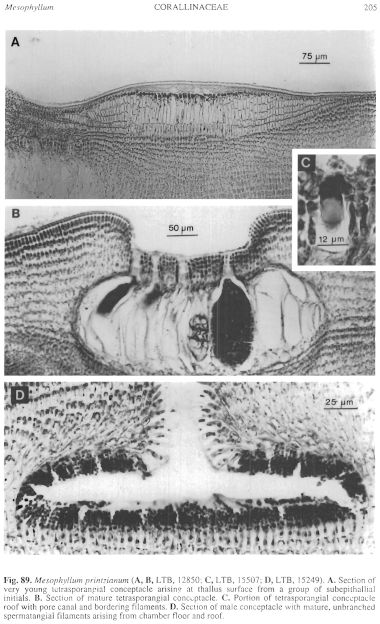
Figure 89 enlarge
Fig. 89. Mesophyllum printzianum (A, B, LTB, 12850; C, LTB, 15507; D, LTB, 15249). A. Section of very young tetrasporangial conceptacle arising at thallus surface from a group of subepithallial initials. B. Section of mature tetrasporangial conceptacle. C. Portion of tetrasporangial conceptacle roof with pore canal and bordering filaments. D. Section of male conceptacle with mature, unbranched spermatangial filaments arising from chamber floor and roof.

|
Email Contact: State Herbarium of South Australia |

|


