|
|
|
|
|
|||||||||||
|
Electronic Flora of South Australia Species Fact Sheet
Phylum Rhodophyta – Class Florideophyceae – Order Rhodymeniales – Family Champiaceae
Selected citations: Guiler 1952: 94. Lucas & Perrin 1947: 207. May 1965: 362. Reedman & Womersley 1976: 81, figs 2E, F, 11A.
Thallus (Fig. 50A) medium to dark red-brown, 6–18 cm high, with one to several main axes with mostly opposite laterals (occasionally one or three from the segment), lower laterals similarly branched. Axes and main laterals 3–5 mm broad, slightly to moderately compressed, laterals with a slender stalk and a rounded apex; diaphragms evident throughout thallus, 2–3 (–4) mm apart in older parts, 1–2 mm apart in branches, branches slightly constricted at diaphragms. Holdfast discoid to slightly lobed, 2–6 mm across; epilithic. Structure multiaxial with peripheral and central initials, developing a single-layered cortex of compact polygonal cells 35–50 µm across, cutting off 1–3 small cells from their outer corners and more numerous such cells near the thallus base; longitudinal filaments both peripheral and central with one or two cells between the diaphragms, usually each cell bearing a secretory cell; diaphragms monostromatic, cells angular, elongate in the compressed branches. Old branches with cortical cells differing on the two sides (Fig. 50B, C) and the space between the diaphragms becoming filled with rhizoids. Rhodoplasts discoid to elongate, ribbon like and branched in inner cells.
Reproduction: Gametangial thalli dioecious. Early female reproduction unknown. Carposporophyte branched, with sterile gonimoblast filaments bearing terminal ovoid carposporangia 30–60 µm in diameter. Cystocarps (Fig. 50D) scattered, conical to urceolate, 0.7–1.3 mm in diameter, pericarp 100–300 µm and 6–12 cells thick, ostiolate. Spermatangia (Fig. 51A) in sori on either side of the diaphragms of lesser branches, cut off from chains of initials from the small cortical cells, ovoid and 3–6 µm in diameter.
Tetrasporangia (Fig. 50E) scattered in lesser branches, transformed from cortical cells and protruding inwards (Fig. 51B), 80–100 µm in diameter, tetrahedrally divided.
Type from Sandy Bay, R. Derwent Estuary, Tas. (Lucas, Oct. 1925); lectotype in Herb. Lucas, NSW, 136559, isolectotypes in NSW 136558 and AD, Al2237.
Selected specimens: Bicheno, Tas., 6 m deep at Cod Rock (Brown, McGeary & Womersley, 5.xi.1982; AD, A55565). Browns R., Tas. (Lucas, Oct. 1923; NSW, 136562). Snug, Tas. (Lucas, Aug. 1925; NSW, 136560).
Distribution: SE Tasmania.
Taxonomic notes: C. insignis is similar to C. viridis in having central as well as peripheral longitudinal filaments, but differs in the broader, compressed branches which are mostly distichously branched and have longer, slender, bases.
References:
GUILER, E.R. (1952). The marine algae of Tasmania. Check List with localities. Pap. Proc. R. Soc. Tasm. 86, 71–106.
LUCAS, A.H.S. & PERRIN, F. (1947). The Seaweeds of South Australia. Part 2. The Red Seaweeds. (Govt Printer: Adelaide.)
MAY, V. (1965). A census and key to the species of Rhodophyceae (red algae) recorded from Australia. Contr. N.S.W. natn. Herb. 3, 349–429.
REEDMAN, D.J. & WOMERSLEY, H.B.S. (1976). Southern Australian species of Champia and Chylocladia (Rhodyméniales: Rhodophyta). Trans. R. Soc. S. Aust. 100, 75–104.
The Marine Benthic Flora of Southern Australia Part IIIB complete list of references.
Publication:
Womersley, H.B.S. (28 June, 1996)
The Marine Benthic Flora of Southern Australia
Rhodophyta. Part IIIB. Gracilarialse, Rhodymeniales, Corallinales and Bonnemaisoniales
Reproduced with permission from The Marine Benthic Flora of Southern Australia Part IIIB 1996, by H.B.S. Womersley. Australian Biological Resources Study, Canberra. Copyright Commonwealth of Australia.
Illustrations in Womersley Part IIIA, 1996: FIGS 50, 51A, B.
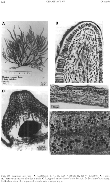
Figure 50 enlarge
Fig. 50. Champia insignis. (A, Lectotype; B, C, E, AD, A55565; D, NSW, 136559). A. Habit. B. Transverse section of older branch. C. Longitudinal section of older branch. D. Section of cystocarp. E. Surface view of compressed branch with tetrasporangia.
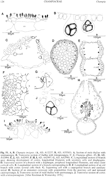
Figure 51 enlarge
Fig. 51. A, B. Champia insignis. (A, AD, Al2237; B, AD, A55565). A. Section of male thallus with spermatangia. B. Transverse section of thallus with tetrasporangia. C–J. Champia affinis. (C, D, AD, A42994; E, J, AD, A42993; F, H, I, AD, A42997; G, AD, A42990). C. Longitudinal section of branch apex showing development of cortex, longitudinal filaments with secretory cells and diaphragms. D. Transverse section of a branch with a diaphragm, peripheral longitudinal filaments, and small outer cortical cells. E. Longitudinal section of a branch corresponding to D. F. Longitudinal section of an old branch with cortex several cells thick. G. Supporting cell with a carpogonial branch and auxiliary initial. H. Section of an immature cystocarp with gonimoblast from the auxiliary cell and terminal carposporangia. I. Transverse section of male thallus with spermatangia. J. Transverse section of cortex with a tetrasporangium. [After Reedman & Womersley 1976.]

|
Email Contact: State Herbarium of South Australia |

|


