|
|
|
|
|
|||||||||||
|
Electronic Flora of South Australia Species Fact Sheet
Phylum Rhodophyta – Class Florideophyceae – Order Nemaliales – Family Galaxauraceae
Selected citations: Huisman 1986: 282, figs 36–47.
Thallus (Fig. 27G) medium red to red-brown, 3–10 cm high, subdichotomously branched every (2–) 5–20 mm, constricted or not at the branchings, branches 0.5–2 mm in diameter, cylindrical, with bluntly pointed apices. Holdfast discoid, 1–2 mm across; epilithic. Structure multiaxial, developing a central core of slender, branched, entwined filaments 2–4 (–8) µm in diameter, from which radiate dichotomous medullary filaments terminating in 2–3 ovoid rhodoplastic cells 8–12 µm in diameter, with an outer layer (Fig. 27H) of colourless utricles, polygonal in surface view, 25–35 µm long and 18–25 µm in diameter.
Reproduction: Sexual thalli monoecious. Monosporangia cut off from elongate rhodoplastic cells penetrating between the hypodermal cells. Carpogonial branches (Fig. 28E) 3-celled, developing on outer medullary filaments, with the hypogynous cell cutting off two sterile branches, 1-celled and 2-celled, and the basal cell producing 3–4 sterile branches which form the involucre after fertilization. Fertilized carpogonium cutting off several initials forming branched gonimoblast filaments (Fig. 28F) with chains of ovoid to clavate carposporangia 10–15 µm long and 4–7 µm in diameter; fusion cell present. Cystocarps (Fig. 27I) 200–300 µm in diameter, ostiolate, with a well developed involucre. Spermatangial filaments scattered or in dense sori, arising from hypodermal cells and penetrating between the utricles, with terminal ovoid spermatangia 2–4 µm in diameter.
Type from Tsinglan-Kang, Wenchang, Hainan, China (Tseng 792, 7.iv.1934); in Herb. Tseng.
Selected specimens: (see also Huisman 1986, p. 282): Pearson I., S. Aust., 28 m deep (Shepherd, 9.i.1969; AD, A34001). Snapper Rock, Port Lincoln, S. Aust., 7–8 m deep (Baldock, 1.i.1964; AD, A27082). Marino, S. Aust., drift (Womersley, 26.x.1975; AD, A46641). Muston, American R. inlet,
Distribution: China.
On all Australian coasts with moderate to slight water movement, usually 3–30 m deep.
Taxonomic notes: Kangaroo I., S. Aust., 4–5 m deep (Shepherd, 29.xii.1977; AD, A48963). Cape Northumberland, S. Aust., 15 m deep 1.3 km off shore (Shepherd, 1.ii.1978; AD, A55039). Lonsdale Bay, Vic., 6–7 m deep (Huisman & O'Brien, 20.x.1981; MELU, A24299). Popes Eye, Port Phillip, Vic., 18 m deep (Millar & Huisman, 20.iii.1981; MELU, 23965). Marion Bay, Tas., 6 m deep (Shepherd, 13.ii.1970; AD, A35633).
References:
HUISMAN, J.M. (1986). The red algal genus Scinaia (Galaxauraceae, Nemaliales) from Australia. Phycologia 25, 271–296.
TSENG, C.K. (1941). Studies on the Chaetangiaceae of China. Bull. Fan Mem. Inst. Biol., Bot. Ser. 11, 83–116, Plates 8–10.
TSENG, C.K. (1983). Common seaweeds of China. (Science Press: Beijing.)
The Marine Benthic Flora of Southern Australia Part IIIA complete list of references.
Publication:
Womersley, H.B.S. (14 January, 1994)
The Marine Benthic Flora of Southern Australia
Rhodophyta. Part IIIA, Bangiophyceae and Florideophyceae (to Gigartinales)
Reproduced with permission from The Marine Benthic Flora of Southern Australia Part IIIA 1994, by H.B.S. Womersley. Australian Biological Resources Study, Canberra. Copyright Commonwealth of Australia.
Illustrations in Womersley Part IIIA, 1994: FIGS 27 G–I, 28E, F.
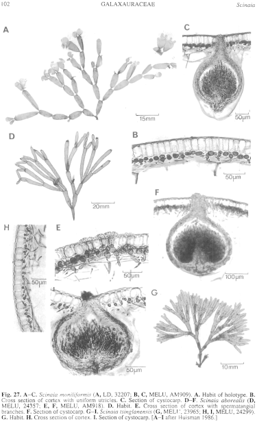
Figure 27 enlarge
Fig. 27. A–C. Scinaia moniliformis (A, LD, 32207; B, C, MELU, AM909). A. Habit of holotype. B. Cross section of cortex with uniform utricles. C. Section of cystocarp. D–F. Scinaia aborealis (D, MELU, 24357; E, F, MELU, AM918). D. Habit. E. Cross section of cortex with spermatangial branches. F. Section of cystocarp. G–I. Scinaia tsinglanensis (G, MELU, 23965; H, I, MELU, 24299). G. Habit. H. Cross section of cortex. I. Section of cystocarp. [A–I after Huisman 1986.]
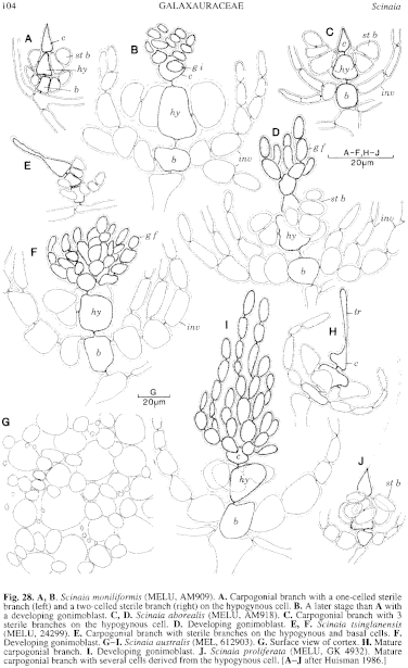
Figure 28 enlarge
Fig. 28. A, B. Scinaia moniliformis (MELU, AM909). A. Carpogonial branch with a one-celled sterile branch (left) and a two-celled sterile branch (right) on the hypogynous cell. B. A later stage than A with a developing gonimoblast. C, D. Scinaia aborealis (MELU, AM918). C. Carpogonial branch with 3 sterile branches on the hypogynous cell. D. Developing gonimoblast. E, F. Scinaia tsinglanensis (MELU, 24299). E. Carpogonial branch with sterile branches on the hypogynous and basal cells. F. Developing gonimoblast. G–I. Scinaia australis (MEL, 612903). G. Surface view of cortex. H. Mature carpogonial branch. I. Developing gonimoblast. J. Scinaia proliferata (MELU, GK 4932). Mature carpogonial branch with several cells derived from the hypogynous cell. [A–J after Huisman 1986.]

|
Email Contact: State Herbarium of South Australia |

|


