|
|
|
|
|
|||||||||||
|
Electronic Flora of South Australia Species Fact Sheet
Phylum Rhodophyta – Class Florideophyceae – Order Gigartinales – Family Acrotylaceae
Selected citations: Harvey 1859a: pl. 99; 1859b: 317. Kraft 1977a: 101, figs 1, 2, 3A–C, 10, 11, 13D. Kützing 1869: 13, pl. 36a, b. Kylin 1932: 68, figs 20A, B, 21B. Lucas & Perrin 1947: 146, fig. 20. Millar & Kraft 1993: 15.
Synonym
Acrotylus australis var. constricta Sonder ex Kützing 1869: 13, pl. 36C.
Thallus (Fig. 120A) dark red-brown, cartilaginous, 5–20 cm high, subdichotomous and largely complanately branched but with short lateral branches from the margins (Fig. 121A), especially in the mid thallus, branches linear, compressed, 1–3 mm broad, often constricted, apices rounded. Holdfast crustose, 2–5 mm across, with one to several fronds; epilithic or epiphytic on larger brown or red algae. Structure multiaxial (Fig. 121C), developing a broad, densely filamentous medulla and a pseudoparenchymatous cortex 5–8 cells thick, cells of the inner two layers (Fig. 121C) somewhat separated, elongate-ovoid to clavate and 10–16 (–20) µm in diameter, outer cells ovoid, 2–3 µm in diameter. Rhodoplasts discoid to elongate, few per cell.
Reproduction: Sexual thalli monoecious; procarpic. Carpogonial branches (Fig. 121D) 3 (–5) on an inner cortical (supporting) cell, 3-celled, sometimes with a sterile cell on the basal cell, inwardly directed with reflexed trichogynes. Supporting cell becoming the auxiliary cell, following diploidization cutting off several subspherical gonimoblast initials (Fig.
121E) which enter the surrounding layer of nutritive tissue formed from adjacent inner cortical cells; gonimoblast and nutritive envelope expanding as the auxiliary cell breaks down, producing a central cavity lined by centripetal gonimoblast filaments which produce tufts bearing terminal, subspherical to ovoid carposporangia 12–18 µm in diameter. Cystocarps (Figs 120B, 121A) deeply embedded, 0.7–1 mm across, with filamentous enveloping tissue and an ostiole developed by rupture of the cortex. Spermatangia (Fig.
121F)in clusters in the cortex of small laterals, with 3–6 initials each forming 2 ovoid spermatangia 2–3 µm in diameter.
Tetrasporangia (Fig. 121G) in slightly raised nemathecia (Fig. 121B), mainly near branch tips, basally pit-connected to mid cortical cells, ovoid, 45–60 µm long and 15–20 µm in diameter, zonately divided.
Type from Sydney, N.S.W.; holotype in Herb. Agardh, LD, 32611.
Selected specimens: Elliston, S. Aust., 20 m deep outside bar (Shepherd, 14.v.1971; AD, A38689). Wanna, S. Aust., drift (Warnersley, 19.ii.1959; AD, A22365). Brown Beach, Yorke Pen., S. Aust., 6–12 m deep (Shepherd, 14.iv.1963; AD, A26560). Pennington Bay, Kangaroo I., S. Aust., drift (Womersley, 27.i.1946; AD, A2919). Port Elliot, S. Aust., drift (Womersley, 23.v.1953; AD, A18736). Cape Jaffa, S. Aust., drift (Womersley, 31.viii.1949; AD, A10882). Robe, S. Aust., drift (Warnersley, 29.viii.1949; AD, A11020). Cape Lannes, S. Aust., drift on Acrocarpia (G. & C. Kraft, 16.i.1971; AD, A44746; MELU, K3209). Stinky Bay, Nora Creina, S. Aust., drift (G. & C. Kraft, 16.xii.1970; AD, A44747). Point Roadknight, Vic., drift (Sinkora A1441, 29.xi.1971; AD, A43182). Point Lonsdale, Vic., 3–5 m deep on Acrocarpia (Kraft, 20.i.1975; MELU, K5126). Walkerville, Vic., drift (Sinkora A2099, 27.ii.1975; AD, A48398). Musselroe Bay, Tas., drift (Perrin, Aug. 1940; AD, A8457).
Distribution: Elliston, S. Aust., to Sydney, N.S.W., and around Tasmania.
Taxonomic notes: Acrotylus australis is common on rough-water coasts, usually in deep water or shaded situations. It is superficially similar to Peltasta australis (Dicranemataceae) and Adelophycus corneus (Nemastomataceae), differing from the former by its broad filamentous medulla and lack of a stoloniferous base, and from the latter by the sharp separation of cortex and medulla and by lack of cortical gland cells; reproductively these taxa are quite distinct. Acrotylus is most commonly epiphytic on Acrocarpia but also occurs on various red algae (e.g. Gelidium, Pterocladia, Laurencia, Cladurus).
References:
AGARDH, J.G. (1849). Algologiska bidrag. Öfvers. K. Svenska Vetensk. Akad. Rh-h. 6(3), 79–89.
AGARDH, J.G. (1851). Species Genera et Ordines Algarum. Vol. 2, Part 1, 1–336 + index. (Gleerup: Lund.)
AGARDH, J.G. (1876). Species Genera et Ordines Algarum. Vol. 3, Part 1 - Epicrisis systematis Floridearum, pp. i-vii, 1–724. (Weigel: Leipzig.)
AGARDH, J.G. (1879). Florideernes morphologi. K. Svenska Vetensk. Akad. Handl. 15(6), 1–199, Plates 1–33.
HARVEY, W.H. (1859a). Phycologia Australica. Vol. 2, Plates 61–120. (Reeve: London.)
HARVEY, W.H. (1859b). Algae. In Hooker, J.D., The Botany of the Antarctic Voyage. Flora Tasmaniae. Vol. II, pp. 282–320.
KÜTZING, F.T. (1869). Tabulae Phycologicae. Vol. 19. (Nordhausen.)
KRAFT, G.T. (1977a). Studies of marine algae in the lesser-known families of the Gigartinales (Rhodophyta). I. The Acrotylaceae. Aust. J. Bot. 25, 97–140.
KYLIN, H. (1932). Die Florideenordnung Gigartinales. Lunds Univ. Årsskr. N.F. Avd. 2, 28 (8), 1–88, Plates 1–28.
LUCAS, A.H.S. & PERRIN, F. (1947). The Seaweeds of South Australia. Part 2. The Red Seaweeds. (Govt Printer: Adelaide.)
MILLAR, A.J.K. & KRAFT, G.T. (1993). Catalogue of Marine and Freshwater Red Algae (Rhodophyta) of New South Wales, including Lord Howe Island, South-western Pacific. Aust. Syst. Bot. 6, 1–90.
The Marine Benthic Flora of Southern Australia Part IIIA complete list of references.
Publication:
Womersley, H.B.S. (14 January, 1994)
The Marine Benthic Flora of Southern Australia
Rhodophyta. Part IIIA, Bangiophyceae and Florideophyceae (to Gigartinales)
Reproduced with permission from The Marine Benthic Flora of Southern Australia Part IIIA 1994, by H.B.S. Womersley. Australian Biological Resources Study, Canberra. Copyright Commonwealth of Australia.
Illustrations in Womersley Part IIIA, 1994: FIGS 120A, B, 121 A–G.
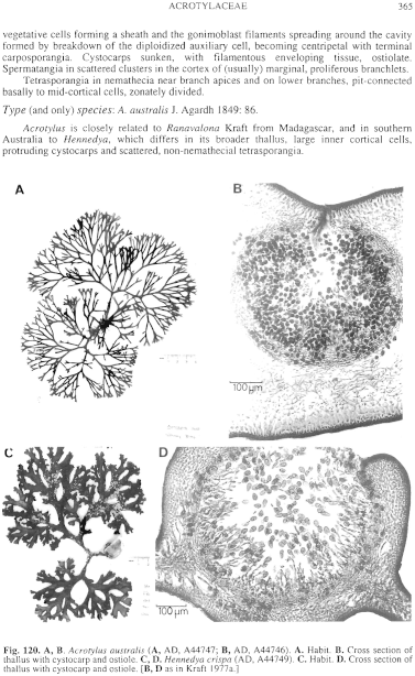
Figure 120 enlarge
Fig. 120. A, B. Acrotylus australis (A, AD, A44747; B, AD, A44746). A. Habit. B. Cross section of thallus with cystocarp and ostiole. C, D. Hennedya crispa (AD, A44749). C. Habit. D. Cross section of thallus with cystocarp and ostiole. [B, D as in Kraft 1977a.]
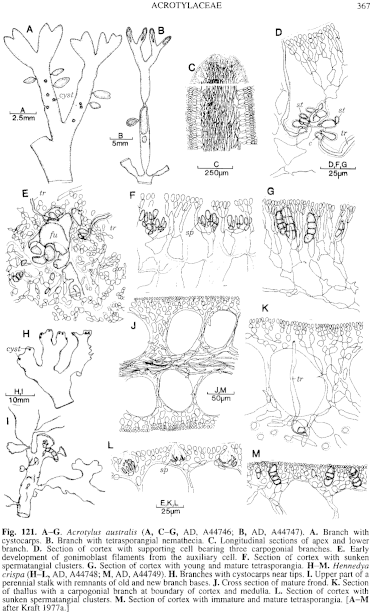
Figure 121 enlarge
Fig. 121. A–G. Acrotylus australis (A, C–G, AD, A44746; B, AD, A44747). A. Branch with cystocarps. B. Branch with tetrasporangial nemathecia. C. Longitudinal sections of apex and lower branch. D. Section of cortex with supporting cell bearing three carpogonial branches. E. Early development of gonimoblast filaments from the auxiliary cell. F. Section of cortex with sunken spermatangial clusters. G. Section of cortex with young and mature tetrasporangia. H–M. Hennedya crispa (H–L, AD, A44748; M, AD, A44749). H. Branches with cystocarps near tips. I. Upper part of a perennial stalk with remnants of old and new branch bases. J. Cross section of mature frond. K. Section of thallus with a carpogonial branch at boundary of cortex and medulla. L. Section of cortex with sunken spermatangial clusters. M. Section of cortex with immature and mature tetrasporangia. [A–M after Kraft 1977a.]

|
Email Contact: State Herbarium of South Australia |

|


