|
|
|
|
|
|||||||||||
|
Electronic Flora of South Australia Species Fact Sheet
Phylum Phaeophyta – Order Chordariales – Family Scytothamnaceae
Selected citations: Asensi 1975: 269, figs 1–4. Clayton 1986: 371, figs 6,11–34. Lindauer et al. 1961: 268, fig. 70. Nelson & Adams 1983: 87, fig. 12. Pedersen 1984: 64. Skottsberg 1921: 34, fig. 14 a,b. Womersley 1967: 247.
Synonym
Dictyosiphon fasciculatus Hooker & Harvey in Harvey & Hooker 1845: 178, pl. 69 fig. 1.
Thallus (Fig. 42B, 43E) medium to dark brown, much branched with several to many axes with 1–3 orders of irregularly arranged, terete, hollow branches, with older branches becoming slightly compressed, 5–15 cm long with axes (300–) 500–2000 µm (-3 mm) in diameter, decreasing to slender ultimate branches (100–) 200–500 µ in diameter, tapering to a pointed to rounded apex and slightly constricted base; epilithic, attached by a discoid holdfast (1–) 2–3 (–4) mm across. Medulla multiaxial from a group of apical cells, producing haplostichous longitudinal filaments which differentiate into an outer medulla (Fig. 43F) of closely appressed longitudinal filaments 8–12 µm in diameter with cells L/B 4–10 and an inner medulla of looser filaments and hyphae 2–4 µm in diameter, with the filaments separating in older parts and the medulla becoming hollow but usually traversed by filaments. Cortex (Fig. 43F,G) dense, 20–45 µm thick, of radial rows of 3–5 more or less isodiametric (to L/B 2) cells 6–10 µm in diameter, branched and fairly closely associated laterally, with the outer layers appearing pseudoparenchymatous. Phaeoplasts (Fig. 43H) single and stellate with a central pyrenoid, becoming elongate-discoid and several per cell (Asensi 1975, fig. 1A,M). Phaeophycean hairs grouped in shallow cryptostomata, 4–6 µm in diameter.
Microthallus gametophytic, filamentous, branched.
Reproduction: Reproduction by ovoid to globose unilocular meiosporangia (Fig. 43F,G) lying within the cortex and visible in surface view, formed from young cortical cells and reaching the thallus surface, 30–50 µm long and 20–30 (–35) µ in diameter. Gametophytes dioecious, producing isogametes (Clayton 1986).
Type from the Auckland Is; in BM (ex K).
Distribution: Subantarctic islands; Chile; New Zealand from Cook Strait south. In southern Australia, only known from south-eastern Tasmania.
Taxonomic notes: Scytothamnus fasciculatus is only known in Australia from the above collections, but can be locally common. At the Tesselated Pavement it occurred as dense patches at the same level as Scytosiphon. It agrees well with New Zealand specimens of this species, differing from S. australis largely in robustness of the thallus.
Pedersen (1984, p. 64) considers that S. fasciculatus should be provisionally placed in Dictyosiphon because of longitudinal divisions in the young macrothallus; further detailed studies are clearly needed.
References:
ASENSI, A.O. (1975). La estructura, la distribucion y el cultivo de Scytothamnus fasciculatus (Hook. et Harv.) Cotton (Phaeophyta). Physis (B. Aires) A, 34, 269–282.
CLAYTON, M.N. (1986). Culture studies on the life history of Scytothamnus australis and Scytothamnus fasciculatus (Phaeophyta) with electron microscope observations on sporogenesis and gametogenesis. Br. phycol. J. 21, 371–386.
COTTON, A.D. (1915). Cryptogams from the Falkland Islands collected by Mrs. Vallentin. J. Linn. Soc. (Bot.) 43, 137–231, Plates 4–10.
HARVEY, W.H. & HOOKER, J.D. (1845). The Botany of the Antarctic Voyage of H.M. Discovery Ships Erebus and Terror in the years 1839–1843. I. Flora Antarctica. Part I. Algae, pp. 175–193, Plates 69–78.
LINDAUER, V.W., CHAPMAN, V.J. & AIKEN, M. (1961). The marine algae of New Zealand. II. Phaeophyceae. Nova Hedwigia 3, 129–350, Plates 57–97.
NELSON, W.A. & ADAMS, N.M. (1983). A taxonomic revision of the families Chordariaceae and Chordariopsidaceae (Phaeophyta) in New Zealand. N.Z. J. Bot. 21, 77–92.
PEDERSEN, P.M. (1984). Studies on primitive brown algae (Fucophyceae). Opera Bot. 74, 1–76.
SKOTTSBERG, C. (1921). Botanische Ergebnisse der Schwedischen Expedition nach Patagonien und dem Feuerlande, 1907–1909. VIII. Marine Algae. I. Phaeophyceae. K. Svenska Vetenskapsakad. Handl. 61(11), 1–56.
WOMERSLEY, H.B.S. (1967). A critical survey of the marine algae of southern Australia. II. Phaeophyta. Aust. J. Bot. 15, 189–270.
The Marine Benthic Flora of Southern Australia Part II complete list of references.
Publication:
Womersley, H.B.S. (14 December, 1987)
The Marine Benthic Flora of Southern Australia
Part II
©Board of the Botanic Gardens and State Herbarium, Government of South Australia
Illustrations in Womersley Part II, 1997: FIGS 42B, 43 E–H.
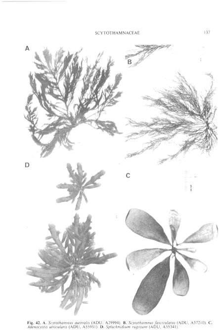
Figure 42 enlarge
Fig. 42. A. Scytothamnus australis (ADU, A29994). B. Scytothamnus fasciculatus (ADU, A57210). C. Adenocystis utricularis (ADU, A55951). D. Splachnidium rugosum (ADU, A55341).
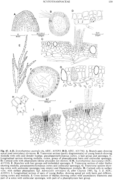
Figure 43 enlarge
Fig. 43. A–D. Scytothamnus australis (A, ADU, A55293; B–D, ADU, A31756). A. Branch apex showing apical (and intercalary) divisions. B. Transverse section (partly diagrammatic) of young branch showing medulla with cells and slender hyphae, pseudoparenchymatous cortex, a hair group and sporangia. C. Longitudinal section showing medulla, cortex, group of phaeophycean hairs and unilocular sporangia. D. Cortical cells with phaeoplasts (dense physodes not shown). E–H. Scytothamnus fasciculatus (ADU, A57210). E. Branches with hair groups and embedded sporangia. F. Transverse section of older thallus showing medulla, pseudoparenchymatous cortex and unilocular sporangia. G. Transverse section showing cortex with unilocular sporangia (one empty) and filamentous medulla. H. Cortical cells in surface view, with stellate phaeoplasts. I,J. Adenocystis utricularis (I, after Clayton 1985, fig. 3; J. ADU, A55951). I. Longitudinal section of apex of young thallus, showing apical pit with hairs and differentiating cortex and medulla. J. Cross section of thallus showing cortex with assimilatory filaments and part of a sorus with unilocular sporangia, with part of a phaeophycean hair group.

|
Email Contact: State Herbarium of South Australia |

|


