|
|
|
|
|
|||||||||||
|
Electronic Flora of South Australia Species Fact Sheet
Phylum Phaeophyta – Order Sporochnales
Selected citations: Womersley 1967: 239.
Thallus (Fig. 97C) medium brown, 15–20 cm long, much branched irregularly radially with long and short laterals intermixed, each branch with a prominent tuft of assimilatory filaments 2–5 mm long (Fig. 98D), with a small rhizoidal holdfast 1–2 mm across; probably epilithic. Growth apical with a convex branch meristem surmounted by the trichothallic assimilatory filaments, each with a meristem 3–5 cells above their base, 20–35 µm in diameter with cells L/B (2–) 3–5 above. New tufts of filaments and laterals arise on any part of the branches. Fronds (Fig. 98D) terete, laterals 2–10 mm apart, 0.5–1 mm in diameter below, tapering to 200–300 µm below apices. Structure haplostichous and pseudoparenchymatous, with a medulla of elongate cells, becoming small-celled outwardly, and bearing a continuous cortex of papillate filaments (Fig. 98E) mostly 2 cells long, with the terminal cell distinctly larger, more or less obovoid, 20–40 µm long and 16–36 µm in diameter; papillate cells and outer medullary cells phaeoplastic.
Reproduction: Reproduction by clavate to elongate-ovoid unilocular sporangia (Fig. 98E) scattered over the thallus between the papillate filaments, sessile or with a short basal cell, 20–30 µm long and (8–) 10–15 µm in diameter. Gametophyte unknown.
Type from Port Phillip Heads, Vic. (Wilson, 3.i.1889); in LD, lectotype 50041.
Distribution: Only known from the type and other Port Phillip Heads, Vic., specimens in LD and MEL (Wilson, 3.i.1889; MEL, 15885; 3.ii.1890; MEL, 15888; 9.ii.1894; MEL, 15887; 12.i.1895; MEL, 15889).
Taxonomic notes: N. lophocladia has not been found in southern Australia since the original collections of J.B. Wilson. As J. Agardh commented, it is closely related to N. filiformis from the Mediterranean, and detailed comparisons are needed as to whether the Australian plant is distinct or a chance introduction of the Mediterranean species which has not persisted. J. Agardh's description may be considered to be invalid as he was doubtful about the species, but it is recognised here pending critical comparisons with N. filiformis.
References:
AGARDH, J.G. (1897). Analecta Algologica. Cont. IV. Ada Univ. lund. 33, 1–106, Plates 1, 2.
WOMERSLEY, H.B.S. (1967). A critical survey of the marine algae of southern Australia. II. Phaeophyta. Aust. J. Bot. 15, 189–270.
The Marine Benthic Flora of Southern Australia Part II complete list of references.
Publication:
Womersley, H.B.S. (14 December, 1987)
The Marine Benthic Flora of Southern Australia
Part II
©Board of the Botanic Gardens and State Herbarium, Government of South Australia
Illustrations in Womersley Part II, 1997: FIGS 97C, 98D,E.
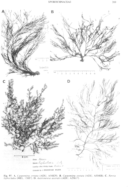
Figure 97 enlarge
Fig. 97. A. Carpomitra costata (ADU, A54829). B. Carpomitra costata (ADU, A55408). C. Nereia lophocladia (MEL, 15887). D. Austronereia australis (ADU, A29617).
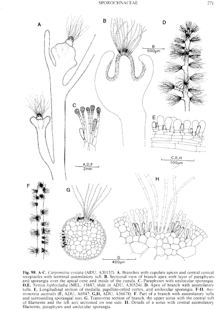
Figure 98 enlarge
Fig. 98. A–C. Carpomitra costata (ADU, A30152). A. Branches with cupulate apices and central conical receptacles with terminal assimilatory tuft. B. Sectional view of branch apex with layer of paraphyses and sporangia over the apical cone and inside of the cupula. C. Paraphyses with unilocular sporangia. D,E. Nereia lophocladia (MEL, 15887, slide in ADU, A30524). D. Apex of branch with assimilatory tufts. E. Longitudinal section of medulla, papillate-celled cortex, and unilocular sporangia. F–H. Austronereia australis ( F, ADU, A6947; G,H, ADU, A56678). F. Part of a branch with assimilatory tufts and surrounding sporangial sofi. G. Transverse section of branch, the upper sorus with the central tuft of filaments and the left sori sectioned on one side. H. Details of a sorus with central assimilatory filaments, paraphyses and unilocular sporangia.

|
Email Contact: State Herbarium of South Australia |

|


