|
|
|
|
|
|||||||||||
|
Electronic Flora of South Australia Species Fact Sheet
Phylum Phaeophyta – Order Chordariales – Family Myrionemataceae
Selected citations: Abbott & Hollenberg 1976: 158, fig. 125. Hamel 1935: 88, fig. 24 (1–10). Harvey 1850: pl. 280. Kornmann & Sahling 1983: 46, figs 28, 29. Kylin 1947: 36, fig. 28. Lindauer et al. 1961: 205, fig. 35 (1–7). Loiseaux 1967b: 547, figs 8–11. Womersley 1967: 229.
Thallus (Figs 15A, 16A) light greenish-brown to medium brown, regularly discoid, 0.5–2 (–3) mm across, epiphytic on Ulva (and occasionally on Enteromorpha). Basal layer (Fig. 16B,C) monostromatic, of subdichotomous, radiating, closely adjacent filaments adherent to the host surface (older cells with short attachment pegs), with marginal cells more elongate and inner cells mostly 8–14 µm long, 5–8 broad and 4–8 µm high. Erect assimilatory filaments (Fig. 16C) forming a shallow even dome, arising from almost every cell of the basal layer commencing 10–20 cells within the disc margin, when mature (70–) 100–130 and 5–8 (–10) cells high, with the cells (8–) 15–25 µm long and 4–6 µm in diameter, with the terminal cell slightly clavate, 10–25 µm long and 7–10 µ in diameter; cells with several discoid to irregularly-shaped phaeoplasts, each with 1 (–2) pyrenoids. Hairs phaeophycean, usually with a basal collar, scattered, (6–) 8–12 in diameter.
Reproduction: Plurilocular sporangia on a unicellular pedicel from scattered cells of basal layer, filiform, uniseriate and with 8–12 locules, of similar height to erect filaments (Kylin 1947, fig. 28C–E); not observed on Australian plants. Unilocular sporangia (Fig. 16C) borne laterally on the short basal or second cell of erect filaments, singly or in pairs, ovoid to ovoid-clavate, (25–) 30–60 µm long and 15–20 µm in diameter.
Type from Appin, Scotland; in E.
Selected specimens: Wedge I., S. Aust., on Ulva on rock platform (Baldock, 29.xii.1963; ADU, A27325). Wallaby I., American R. inlet, Kangaroo I., S. Aust., on Ulva, low eulittoral (Womersley, 28.viii.1950; ADU, A15285). Rosetta Bay, Victor Harbor, S. Aust., low eulittoral on U. australis (Womersley, 7.ix.1986; ADU, A57139-"Marine Algae of southern Australia" No. 156a). Encounter Bay, S. Aust., on Enteromorpha compressa, uppermost sublittoral (Skinner, 21.viii.1978; ADU, A49514). Robe, S. Aust., on Ulva, upper sublittoral ( Womersley, 6.xi.1965; ADU, A29639-"Marine Algae of southern Australia" No. 156). Crawfish Rock, Westernport Bay, Vic., on Ulva, lower eulittoral (Womersley, 29.viii.1971; ADU, A39441). Bombay Rock, Tamar Est., Tas., on Ulva, upper sublittoral (Womersley, 27.i.1949; ADU, A10373). The host of M. strangulans is usually Ulva australis.
Distribution: Widespread in temperate seas.
In southern Australia, from Wedge I., S. Aust., to Crawfish Rock, Westernport Bay, Vic. and around Tasmania. Probably more widespread.
Taxonomic notes: Loiseaux (1967b, fig. 11) has shown that European M. strangulans has a heteromorphic life history, with the discoid sporophytes producing either plurilocular neutral sporangia or unilocular sporangia producing zooids which form either a filamentous gametophyte (with isogametes) or which fuse directly. Only unilocular sporangia have been observed on Australian plants and the filamentous gametophytes have not been recorded. Skinner (1981) found a direct life history for southern Australian plants, with the zooids from unilocular sporangia forming new discoid thalli again producing unilocular sporangia. The unilocular sporangia on Australian plants are distinctly more elongate than those figured for European plants.
References:
ABBOTT, I.A. & HOLLENBERG, G.J. (1976). Marine Algae of California. (Stanford Univ. Press: Stanford.)
GREVILLE, R.K. (1827). Scottish Cryptogamic Flora. Vol. 5, Plates 271–300. (Edinburgh.)
HAMEL, G. (1935). Phéophycées de France. Fasc. II, pp. 81–176. (Paris.)
HARVEY, W.H. (1850). Phycologia Britannica. Plates 253–354. (Reeve: London.)
KORNMANN, P. & SAHLING, P.-H. (1983). Meeresalgen von Helgoland: Ergänzung. Helgol. wiss. Meeresunters. 36, 1–65.
KYLIN, H. (1947). Die Phaeophyceen der Schwedischen Westkiiste. Acta Univ. lund. N.F. Avd. 2, 43(4), 1–99, Plates 1–18.
LINDAUER, V.W., CHAPMAN, V.J. & AIKEN, M. (1961). The marine algae of New Zealand. II. Phaeophyceae. Nova Hedwigia 3, 129–350, Plates 57–97.
LOISEAUX, S. (1967b). Recherches sur les cycles de développement des Myrionématacées (Phéophycées). I–II. Hécatonématées et Myrionématées. Rev. Gén. Bot. 74, 529–577, Plates 1, 2.
SKINNER, S. (1981). The taxonomy, morphology and reproduction of the Myrionemaceae, Elachistaceae, Corynophlaeaceae and Giraudyaceae (Phaeophyceae) in southern Australia. Ph. D. thesis, University of Adelaide, unpublished.
WOMERSLEY, H.B.S. (1967). A critical survey of the marine algae of southern Australia. II. Phaeophyta. Aust. J. Bot. 15, 189–270.
The Marine Benthic Flora of Southern Australia Part II complete list of references.
Publication:
Womersley, H.B.S. (14 December, 1987)
The Marine Benthic Flora of Southern Australia
Part II
©Board of the Botanic Gardens and State Herbarium, Government of South Australia
Illustrations in Womersley Part II, 1997: FIGS 15A, 16 A–C.
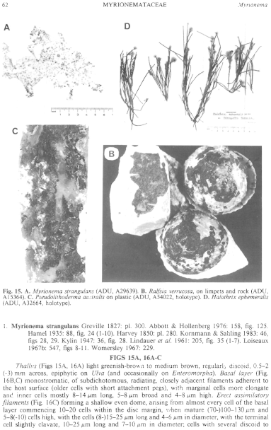
Figure 15 enlarge
Fig. 15. A. Myrionema strangulans (ADU, A29639). B. Ralfsia verrucosa, on limpets and rock (ADU, A15364). C. Pseudolithoderma australis on plastic (ADU, A54022, holotype). D. Halothrix ephemeralis (ADU, A32664, holotype).
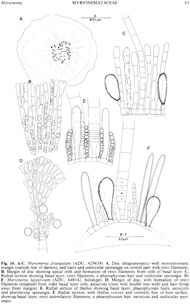
Figure 16 enlarge
Fig. 16. A–C. Myrionema strangulans (ADU, A29639). A. Disc (diagrammatic) with monostromatic margin (outside line of dashes), and hairs and unilocular sporangia on central part with erect filaments. B. Margin of disc showing apical cells and formation of erect filaments from cells of basal layer. C. Radial section showing basal layer, erect filaments, a phaeophycean hair and unilocular sporangia. D–F. Myrionema latipilosum (ADU, A48142, holotype). D. Margin of disc, with formation of erect filaments (stippled) from older basal layer cells, ascocysts (clear with double line wall) and hair (lying away from margin). E. Radial section of thallus showing basal layer, phaeophycean hairs, ascocysts and plurilocular sporangia. F. Radial section, with thallus convex and centrally free of host surface, showing basal layer, erect assimilatory filaments, a phaeophycean hair, ascocysts and unilocular sporangia.

|
Email Contact: State Herbarium of South Australia |

|


