|
|
|
|
|
|||||||||||
|
Electronic Flora of South Australia Species Fact Sheet
Phylum Phaeophyta – Order Chordariales – Family Leathesiaceae
Selected citations: Womersley 1967: 230.
Synonym
Myriactis cystophorae (J. Agardh) Kuckuck 1929: 40, fig. 42.
Thallus (Figs 27B, 28A) dark brown, hemispherical to globose, firm but very mucoid, 2–4 (–6) mm across, epiphytic on Cystophora and other algae. Basal layer of appressed radiating filaments on the host, with no or very slight penetration between the outer host cells, cells isodiametric to slightly elongate, 6–8 µm in diameter. Medulla 200–900 µm high, of erect, branched, closely adjacent filaments arising from every cell of the basal layer, cells ovoid to elongate-ovoid to sub-pyriform below, 20–40 (–50) µm in diameter, becoming smaller above. Determinate cortical filaments (Fig. 28A,B) arising in groups of 1–4 from upper medullary cells, 100–500 µm and (8–) 15–35 (–90) cells long, usually curved above, cells cylindrical below, 6–15 µm in diameter and L/B 3–6, and usually laterally inflated above (globose to often 1:n,1 ,1,11111// 1,,,[11,111/1,,
Reproduction: Plurilocular sporangia (Fig. 28B) borne on slender branch systems from the upper medullary cells, filiform, uniseriate, simple or once branched, 30–50 µm and 8–25 (–36) locules long, 5–7 µm in diameter. Unilocular sporangia (Fig. 28D) often on the same thallus as plurilocular sporangia, borne laterally at base of cortical filaments, ovoid, 50–100 (–160) µm long, 20–30 (–45) µm in diameter.
Type from Port Phillip Heads, Vic. (Harvey, Alg. Aust. Exsicc. 102); holotype in Herb. Agardh, LD, 46193; isotype in NSW.
Selected specimens: Natural Jetty reef, Rottnest I., W. Aust., upper sublittoral on Cystophora brownii (Gordon, 10.xi.1968; ADU, A33330). Point Avoid, S. Aust. in reef pools on C. moniliformis (Womersley, 5.i.1976; ADU, A46767-"Marine Algae of southern Australia" No. 194b). Fishery Bay, S.W. of Port Lincoln, S. Aust., lower eulittoral pools on C. brownii (Skinner, 4.xii.1977; ADU, A48909). Wanna, S. Aust., lower eulittoral pools on Acrocarpia (Skinner, 4.xii.1977; ADU, A48905). Reevesby I., Sir Joseph Banks Group, S. Aust., 0–3.5 m deep on Metagoniolithon (Baldock, 13.xii.1977; ADU, A48923). Balgowan, Yorke Pen., S. Aust., 3–8 m deep on Osmundaria (Kald, 17.xii.1967; ADU, A32188). Aldinga, S. Aust., in upper sublittoral pools on C. polycystidea (Skinner, 11.xi.1977; ADU, A48830-"Marine Algae of southern Australia" No. 194a). Pennington Bay, Kangaroo I., S. Aust., on Sargassum (Womersley, 17.xi.1967; ADU, A32184). Encounter Bay, S. Aust., in lower eulittoral pools on Caulocystis (Skinner, 28.ix.1972; ADU, A48263). Apollo Bay, Vic., in lower eulittoral pools on Xiphophora chondrophylla (Womersley, 23.i.1967; ADU, A31759). Point Lonsdale, Vic., upper sublittoral on Cystophora torulosa (Skinner, 17.i.1979; ADU, A50232). Bicheno, Tas., lower eulittoral pools on C. subfarcinata (Skinner, 22.ii.1978; ADU, A49169).
Distribution: From Rottnest I., W. Aust. around southern Australia and Tasmania to Crookhaven Heads, N.S.W.
Taxonomic notes: 2 3 4 5 fi deltoid on their upper side), sometimes remaining cylindrical, (6–) 8–15 (–20) µm broad or long and L/B 1–1.5 (–2). Phaeoplasts (Fig. 28C) several per cell, discoid to elongate or lobed, each usually with a pyrenoid. Phaeophycean hairs frequent, produced from upper medullary cells, 10–1511m in diameter and greatly exceeding the cortical filaments in length.
New Zealand.
C. cystophorae is common on several species of Cystophora, as well as on a variety of other algae (see above list) along rough-water to moderately sheltered coasts of southern Australia. It varies particularly in the length of the determinate cortical filaments, their upper curvature, and the degree of lateral enlargement of the upper cells. An isotype specimen in NSW shows cortical filaments 10–15 cells long, curved above with considerable lateral enlargement of the upper cells, as described by J. Agardh (1882, p. 22, pl. 1 fig. 1).
New Zealand specimens with long cortical filaments were described by Reinbold (1899b, p. 291) as var. longifila, and elevated to a species by Lindauer et al. [1961, p. 218, fig. 43(5,6)]. The great variation in length [8–60 (–90) cells] of cortical filaments, whether the upper part is curved or not, and in the degree of swelling or lateral enlargement of the cells, include the features ascribed to C. longifila which cannot be separated satisfactorily from C. cystophorae on the characters used by Lindauer et al.
References:
AGARDH, J.G. (1882). Till algernes systematik. Acta Univ. lund. 17, 1–136, Plates 1–3.
KUCKUCK, P. (1929). Fragmente einer Monographie des Pheosporeen. Biol. Anst. Helgol. 17, 1–93.
REINBOLD, T. (1899b). Ergebnisse einer Reise nach dem Pacific. (Prof. Dr. Schauinsland, 1896–1897). Meeresalgen. Abh. Naturwiss. Ver. Bremen 16, 287.
WOMERSLEY, H.B.S. (1967). A critical survey of the marine algae of southern Australia. II. Phaeophyta. Aust. J. Bot. 15, 189–270.
The Marine Benthic Flora of Southern Australia Part II complete list of references.
Publication:
Womersley, H.B.S. (14 December, 1987)
The Marine Benthic Flora of Southern Australia
Part II
©Board of the Botanic Gardens and State Herbarium, Government of South Australia
Illustrations in Womersley Part II, 1997: FIGS 27B, 28 A–D.
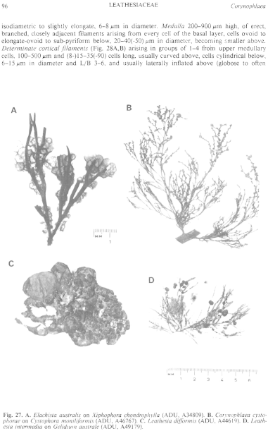
Figure 27 enlarge
Fig. 27. A. Elachista australis on Xiphophora chondrophylla (ADU, A34809). B. Corynophlaea cystophorae on Cystophora tnoniliformis (ADU, A46767). C. Leathesia difformis (ADU, A44619). D. Leathesia intermedia on Gelidium australe (ADU, A49179).
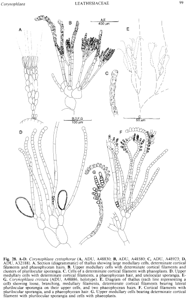
Figure 28 enlarge
Fig. 28. A–D. Corynophlaea cystophorae (A, ADU, A48830; B, ADU, A48580; C, ADU, A48923; D, ADU, A32188). A. Section (diagrammatic) of thallus showing large medullary cells, determinate cortical filaments and phaeophycean hairs. B. Upper medullary cells with determinate cortical filaments and clusters of plurilocular sporangia. C. Cells of a determinate cortical filament with phaeoplasts. D. Upper medullary cells with determinate cortical filaments, a phaeophycean hair, and unilocular sporangia. E–G. Corynophlaea cristata (ADU, A48886, holotype). E. Diagram of thallus (each line representing a cell) showing loose, branching, medullary filaments, determinate cortical filaments bearing lateral plurilocular sporangia on their upper cells, and two phaeophycean hairs. F. Cortical filaments with plurilocular sporangia, and a phaeophycean hair. G. Upper medullary cells bearing determinate cortical filament with plurilocular sporangia and cells with phaeoplasts.

|
Email Contact: State Herbarium of South Australia |

|


