|
|
|
|
|
|||||||||||
|
Electronic Flora of South Australia Species Fact Sheet
Phylum Phaeophyta – Order Chordariales – Family Chordariaceae
Selected citations: Bailey & Womersley in Womersley 1967: 233.
Synonyms
Bactrophora filum (Harvey) J. Agardh 1882: 24, pl. 1 fig. 4.
Castagnea filum (Harvey) Kuckuck 1929: 50, figs 61,62.
Thallus (Fig. 35B) medium to dark brown, very mucoid, simple or with one to a few laterals, 10–50 cm long and (1–) 2–4 (–6) mm in diameter, with a small discoid holdfast 0.5–2 mm across, epiphytic usually on Posidonia or Amphibolis. Basal system of juvenile plants consisting of a pseudoparenchymatous layer of filaments, producing the core of erect medullary filaments. Medulla (Fig. 37A) becoming hollow, of more or less parallel longitudinal cylindrical filaments 25–50 (–80) µm in diameter with cells L/B (1–) 2–4 (–6), with only a few hyphae but with slightly slenderer filaments of shorter cells (L/B 1–2) on the periphery; the latter develop further with age, originating from the outer, larger, medullary filaments or the basal cells of the subcortex. Subcortex slight, 20–60 (–80) µm broad and usually of 1–3 cells (branched if 2 or 3) at right angles to medullary cells, cells ovoid to elongate ovoid, (10–) 20–30 µm in diameter and L/B 1–2. Cortical filaments (Figs 36F, 37A) arising singly or in groups from outer subcortical cells or directly from peripheral medullary cells, simple, determinate, 400–500 (–800) µm and usually 20–30 (–45) cells long, curved to recurved above; upper cells 8–12 µm in diameter and L/B 0.7–1.5 (–2), ovoid or extended on their upper sides, lower cells cylindrical, 6–10 µm in diameter and L/B 4–8. Phaeoplasts (Fig. 36G) several per cell, discoid, each with a pyrenoid; cells with numerous physodes. Phaeophycean hairs (Figs 36F,37A) frequent, arising from outer subcortical cells, (16–) 20–25 (–30) pm in diameter.
Reproduction: Plurilocular sporangia (Fig. 36G) produced from upper cells of curved cortical filaments by successive subdivisions on their upper side to form a cluster 10–20 µm long. Unilocular sporangia (Fig. 37A) on the same or different plants, borne on outer sub-cortical cells, ovoid, 80–100 A m long and 60–80 A m in diameter.
Type from King George Sound, W. Aust. (Harvey, Alg. Aust. Exsicc. 93B); in Herb. Harvey, TCD.
Selected specimens: Safety Bay, W. Aust., on Posidonia australis, drift ( Womersley, 29.ix.1979; ADU, A50740). Tiparra reef, Spencer Gulf, S. Aust., on Posidonia sinuosa, 7 m deep (Shepherd, 9.xii.1980; ADU, A51948-"Marine Algae of southern Australia" No. 251). Cable Hut Bay, Yorke Pen., S. Aust., upper sublittoral on Amphibolis antarctica (Womersley, 9.xi.1980; ADU, A51867). Emu Bay, Kangaroo I., S. Aust., on Posidonia sinuosa, upper sublittoral (Bailey, 15.i.1965; ADU, A32004). Cats Bay, Phillip I., Vic. (Norris, 20.i.1963; ADU, A27488). Wynyard, Tas., on Amphibolis antarctica, upper sublittoral (Gordon, 18.i.1966; ADU, A30013).
Distribution: From Safety Bay, W. Aust. to Twofold Bay, N.S.W. and around Tasmania, on Posidonia spp. and Amphibolis in the sublittoral, rarely on larger algae (e.g. Scaberia), under moderate wave action. It is often common in the upper sublittoral but is recorded to a depth of 26 m.
Taxonomic notes: Cladosiphon filum is one of the more common Chordariaceae, present on the more robust seagrasses during summer months (Sept.-March). It is distinguished by the absence of or only slight branching, the very limited subcortex, the extensive cortex of upwardly curved assimilatory filaments, and the relatively broad phaeophycean hairs. The degree of development of the subcortex, and the length of the cortical filaments, are rather variable. Near the apices, cortical filaments frequently arise directly from peripheral medullary cells, but on older parts further cortical filaments arise from the enlarged basal cell of the cortical filaments, which then becomes a subcortical cell.
References:
AGARDH, J.G. (1882). Till algernes systematik. Acta Univ. lund. 17, 1–136, Plates 1–3.
KUCKUCK, P. (1929). Fragmente einer Monographie des Pheosporeen. Biol. Anst. Helgol. 17, 1–93.
KYLIN, H. (1940). Die Phaeophyceenordnung Chordariales. Acta Univ. lund. N.F. Avd. 2, 36(9), 1–67, Plates 1–8.
WOMERSLEY, H.B.S. (1967). A critical survey of the marine algae of southern Australia. II. Phaeophyta. Aust. J. Bot. 15, 189–270.
The Marine Benthic Flora of Southern Australia Part II complete list of references.
Publication:
Womersley, H.B.S. (14 December, 1987)
The Marine Benthic Flora of Southern Australia
Part II
©Board of the Botanic Gardens and State Herbarium, Government of South Australia
Illustrations in Womersley Part II, 1997: FIGS 35B, 36F,G, 37A.
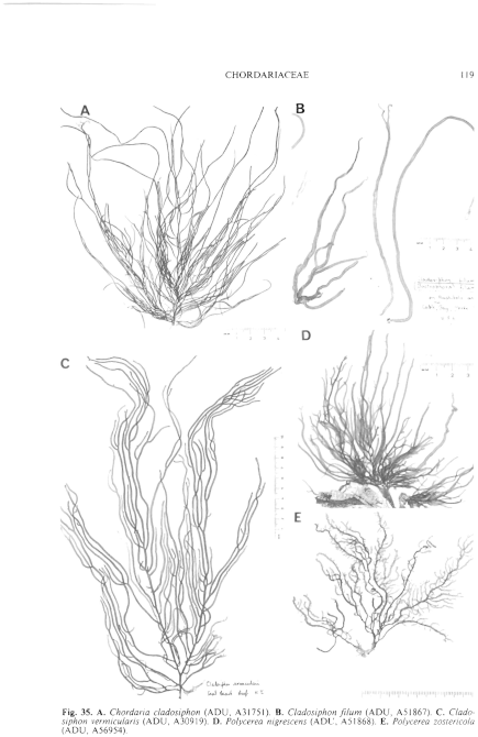
Figure 35 enlarge
Fig. 35. A. Chordaria cladosiphon (ADU, A31751). B. Cladosiphon filum (ADU, A51867). C. Cladosiphon vermicularis (ADU, A30919). D. Polycerea nigrescens (ADU, A51868). E. Polycerea zostericola (ADU, A56954).
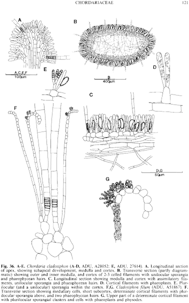
Figure 36 enlarge
Fig. 36. A–E. Chordaria cladosiphon (A–C, ADU, A28052; E, ADU, 27614). A. Longitudinal section of apex, showing subapical development, medulla and cortex. B. Transverse section (partly diagrammatic) showing outer and inner medulla, and cortex of 2-3 celled filaments with unilocular sporangia and phaeophycean hairs. C. Longitudinal section showing medulla and cortex with assimilatory filaments, unilocular sporangia and phaeophycean hairs. D. Cortical filaments with phaeoplasts. E. Plurilocular (and a unilocular) sporangia within the cortex. F,G. Cladosiphon ilium (ADU, A51867). F. Transverse section showing medullary cells, short subcortex, determinate cortical filaments with plurilocular sporangia above, and two phaeophycean hairs. G. Upper part of a determinate cortical filament with plurilocular sporangial clusters and cells with phaeoplasts and physodes.
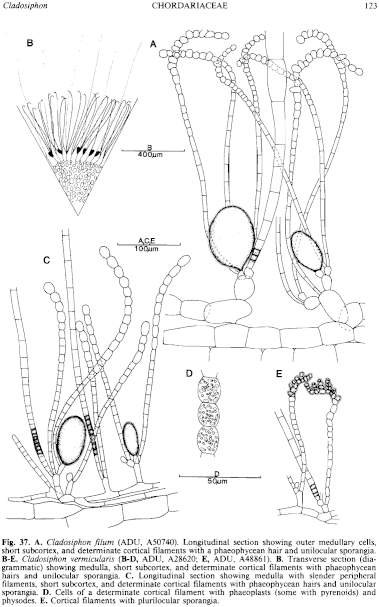
Figure 37 enlarge
Fig. 37. A. Cladosiphon filum (ADU, A50740). Longitudinal section showing outer medullary cells, short subcortex, and determinate cortical filaments with a phaeophycean hair and unilocular sporangia. B–E. Cladosiphon vermicularis (B–D, ADU, A28620; E, ADU, A48861). B. Transverse section (diagrammatic) showing medulla, short subcortex, and determinate cortical filaments with phaeophycean hairs and unilocular sporangia. C. Longitudinal section showing medulla with slender peripheral filaments, short subcortex, and determinate cortical filaments with phaeophycean hairs and unilocular sporangia. D. Cells of a determinate cortical filament with phaeoplasts (some with pyrenoids) and physodes. E. Cortical filaments with plurilocular sporangia.

|
Email Contact: State Herbarium of South Australia |

|


