|
|
|
|
|
|||||||||||
|
Electronic Flora of South Australia Species Fact Sheet
Phylum Rhodophyta – Class Florideophyceae – Order Gigartinales – Family Nemastomataceae
Synonym
Nemastoma feredayae Harvey 1860a: 327, pl. 195A. J. Agardh 1876: 126; 1899: 75. Chapman 1979: 282, fig. 76, pls 95, 96. De Toni 1905: 1663. Fuhrer et al. 1981: pl. 61. Kylin 1932: 7.
Thallus (Fig. 87D) blue-green to brownish (in shallow water plants) to red-brown (in deeper water plants), mucilaginous, 4–35 cm high, subdichotomously and more or less complanately branched at intervals of (2–) 5–20 (–40) mm, often densely branched above, with slightly compressed linear branches (2–) 3–5 mm broad below, tapering to (0.5–) 1–2 (–3) mm broad near the apices, some plants with small proliferations below (or following damage?). Holdfast discoid, 1–5 mm across, bearing one to several fronds; epilithic. Structure of a cortex (Fig. 88A) 150–250 µm thick, of discrete but adjacent branch systems with several basal subdichotomies, cells elongate to ovoid, 4–6 (–8) µm in diameter in shallow water plants to 12–16 (–20) µm in diameter in deeper water plants, and an outer cortex of subdichtomous filaments, the inner cells 4–6 (–10) µm in diameter, L/D 2–4 (–6), tapering to outer unbranched rows of cells (2–) 3–4 µm in diameter and L/D 1–2 (–3). Medulla broad, of longitudinal and entangled filaments 3–6 µm in diameter, with some becoming 10–15 µm in diameter. Rhodoplasts discoid to elongate, few per cell.
Reproduction: Carpogonial branches (Fig. 88A) 3 (–4)-celled with a more or less coiled trichogyne, borne on inner cortical cells. Auxiliary cells (Fig. 88B) in mid cortex below a subdichotomy, following union with a connecting filament producing outwardly a subspherical to conical carposporophyte (Fig. 88C) 120–180 µm across, lying between the cortical filaments; carposporangia ovoid, 8–14 µm in diameter; pericarp and pore absent. Spermatangia unknown.
Tetrasporangia (Fig. 88D) borne on mid cortical cells, elongate-ovoid, 16–30 µm long and 6–16 µm in diameter, cruciately to obliquely divided.
Type from Georgetown, Tas. (Fereday); lectotype in Herb. Harvey, TCD.
Selected specimens: Nuyts Reef, S. Aust., 30 m deep (Shepherd, 26.iii.1980; AD, A52212). Point Sinclair, S. Aust., in shallow pools, rough-water reefs (Womersley, 8.ii.1954; AD, A19622). Vivonne Bay, Kangaroo I., S. Aust., drift (Womersley, 2.i.1949; AD, A10615). Pennington Bay, Kangaroo I., S. Aust., reef edge (Womersley, 5.i.1947; AD, A4341). Robe, S. Aust., 8 m deep inside Baudin Rocks (P. Womersley, 24.viii.1973; AD, A43955). 1.3 km off Middle Point, Cape Northumberland, S. Aust., 14 m deep (Shepherd, 13.iii.1975; AD, A46184). Bridgewater Bay, Vic., upper sublittoral (Womersley, 25.i.1967; AD, A31774). Walkerville, Vic., drift (Sinkora A1638, 23.ii.1972; AD, A43163). Cape Portland, Tas. (Levring, 8.ii.1948; AD, A61269). Safety Cove, Port Arthur, Tas., 7 m deep (Brown & Kenchington, 16.x.1986; AD, A57726). Tasman I., Tas., upper sublittoral (Bennett, 28.vi.1954; AD, A19833). Crayfish Point, Taroona, Tas., 0–6 m deep (Sanderson, 29.v.1992; AD, A61742 -"Marine Algae of southern Australia" No. 369). Dover, Tas., 4–6 m deep under Macrocystis (Kraft 7844 & Sanderson, 8.iv.1988; MELU, 40368–40370). Southport, Tas., upper sublittoral on platform (Wollaston & Mitchell, 27.ii.1964; AD, A27708).
Distribution: New Zealand (North Island).
Nuyts Reef, S. Aust., to Walkerville, Vic., and the N, E and S coasts of Tasmania.
Taxonomic notes: T. feredayae is characterised by its habit, with subdichotomous, complanately branched, branches mostly 3–5 mm broad, but varies in colour, size, density of branching, and dimensions of inner cortical cells depending on the depth. Plants from near low tide level in rough-water conditions are smaller, more densely branched, yellow-brown in colour and have narrow inner cortical cells compared to larger, less branched, red-brown plants from deeper water which usually have ovoid, relatively large, inner cortical cells packed with starch grains.
T. feredayae is similar in habit and dimensions to the type species, T. nakamurae, but differs in that the carpogonial branches are straight instead of bent at right angles and the tetrasporangia are less regularly cruciately divided than in the type species.
References:
AGARDH, J.G. (1876). Species Genera et Ordines Algarum. Vol. 3, Part 1 - Epicrisis systematis Floridearum, pp. i-vii, 1–724. (Weigel: Leipzig.)
AGARDH, J.G. (1899). Analecta Algologica. Cont. V. Acta Univ. lund. 35, 1–160, Plates 1–3.
CHAPMAN, V.J. (1979). The marine algae of New Zealand. Part DI Rhodophyceae. Issue 4: Gigartinales. (Cramer: Germany.)
DE TONI, G.B. (1905). Sylloge Algarum omnium hucusque Cognitarum. Vol. 4. Florideae. Sect. 4, pp. 1523–1973. (Padua.)
FUHRER, B., CHRISTIANSON, I.G., CLAYTON, M.N. & ALLENDER, B.M. (1981). Seaweeds of Australia. (Reed: Sydney.)
HARVEY, W.H. (1860a). Algae. In Hooker, J.D., The Botany of the Antarctic Voyage. 111. Flora Tasmaniae. Vol. II, pp. 321–343, Plates 185–196.
KYLIN, H. (1932). Die Florideenordnung Gigartinales. Lunds Univ. Årsskr. N.F. Avd. 2, 28 (8), 1–88, Plates 1–28.
The Marine Benthic Flora of Southern Australia Part IIIA complete list of references.
Publication:
Womersley, H.B.S. (14 January, 1994)
The Marine Benthic Flora of Southern Australia
Rhodophyta. Part IIIA, Bangiophyceae and Florideophyceae (to Gigartinales)
Reproduced with permission from The Marine Benthic Flora of Southern Australia Part IIIA 1994, by H.B.S. Womersley. Australian Biological Resources Study, Canberra. Copyright Commonwealth of Australia.
Illustrations in Womersley Part IIIA, 1994: FIGS 87D, 88 A–D.
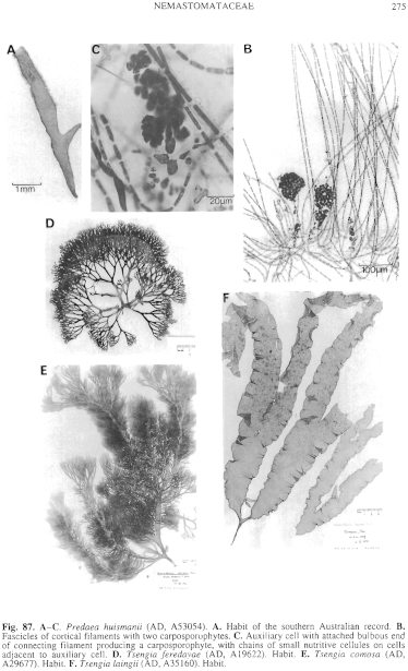
Figure 87 enlarge
Fig. 87. A–C. Predaea huismanii (AD, A53054). A. Habit of the southern Australian record. B. Fascicles of cortical filaments with two carposporophytes. C. Auxiliary cell with attached bulbous end of connecting filament producing a carposporophyte, with chains of small nutritive cellules on cells adjacent to auxiliary cell. D. Tsengia feredayae (AD, A19622). Habit. E. Tsengia comosa (AD, A29677). Habit. F. Tsengia laingii (AD, A35160). Habit.
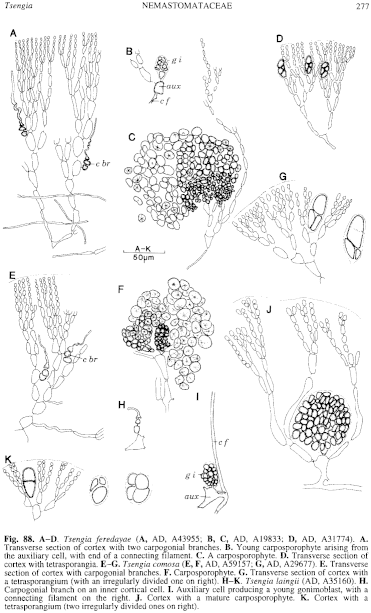
Figure 88 enlarge
Fig. 88. A–D. Tsengia feredayae (A, AD, A43955; B, C, AD, A19833; D, AD, A31774). A. Transverse section of cortex with two carpogonial branches. B. Young carposporophyte arising from the auxiliary cell, with end of a connecting filament. C. A carposporophyte. D. Transverse section of cortex with tetrasporangia. E–G. Tsengia comosa (E, F, AD, A59157; G, AD, A29677). E. Transverse section of cortex with carpogonial branches. F. Carposporophyte. G. Transverse section of cortex with a tetrasporangium (with an irregularly divided one on right). H–K. Tsengia laingii (AD, A35160). H. Carpogonial branch on an inner cortical cell. I. Auxiliary cell producing a young gonimoblast, with a connecting filament on the right. J. Cortex with a mature carposporophyte. K. Cortex with a tetrasporangium (two irregularly divided ones on right).

|
Email Contact: State Herbarium of South Australia |

|


