|
|
|
|
|
|||||||||||
|
Electronic Flora of South Australia Species Fact Sheet
Phylum Rhodophyta – Class Florideophyceae – Order Gigartinales – Family Areschougiaceae
Thallus (Fig. 107D, E) white, darkening on drying, 3–6 mm high, parasitic on Solieria robusta, forming pustules with a basal terete stalk and several blunt to conical lobes 0.5–1 mm long and 300–500 µm in diameter. Structure (Fig. 107F) multiaxial with each axial cell cutting off a single periaxial cell and developing a filamentous medulla of linked cells and a cortex of inner, ovoid to elongate, larger, multinucleate cells 8–14 µm in diameter and an outer cortex of small, ovoid, uninucleate cells 3–6 µm in diameter and L/D 2–3. Endophytic parasite filaments are slender and become pit-connected to cortical cells of the host.
Reproduction: Sexual thalli monoecious; non-procarpic. Carpogonial branches (Fig. 109A) 3-celled, borne on inner cortical cells, directed inwards with a reflexed trichogyne and the hypogynous cell often with a filiform process. Auxiliary cell (Fig. 109B) an inner cortical cell in a darker staining complex, with the fertilized carpogonium producing two unbranched connecting filaments, one cut off by a pit-connection and the other not, connecting to an auxiliary cell which produces inwardly 1 or 2 gonimoblast initials, then developing a fusion cell with radial gonimoblasts forming a surface layer of terminal, ovoid carposporangia 15–20 µm long and 10–15 µm in diameter; carposporophyte (Fig. 107G) with slight enveloping tissue and a central fusion cell with its medullary stalk cell. Cortical filaments adjacent to the carposporophyte form files of cells except just outside the auxiliary cell, resulting in a pit which becomes an ostiole. Spermatangia (Fig. 109C) scattered, either on male lobes or at the base of female lobes, cut off from surface cortical cells, ovoid, about 2 µm in diameter.
Tetrasporangia (Fig. 109D) scattered in the outer cortex, laterally attached, ovoid, 20–30 µm long and 8–15 µm in diameter, zonately divided.
Type from Portsea Pier, Port Phillip Bay, Vic. (Kraft, Huisman & Wynne, 25.iii.1981); holotype in MELU, K 3399.
Selected specimens: Marino, S. Aust., 3–4 m deep (Kraft & Owen, 19.i.1973; MELU, K 4397). Port Noarlunga, S. Aust., 21 m deep (Shepherd, March 1966; AD, A30485). Vivonne Bay, Kangaroo I., S. Aust., 3–6 m deep on jetty piles (Kraft, 15.iv.1973; AD, A43724). Redbanks, Kangaroo I., S. Aust., upper sublittoral (Womersley, 28.xii.1949; AD, Al2868). Off St Leonards, Port Phillip, Vic., 4 m deep (Macpherson, 5.v.1963; AD, A28669). Crawfish Rock, Westernport Bay, Vic., 0 m deep (Watson, 29.v.1974; AD, A44419).
Distribution: Marino, S. Aust., to Westernport Bay, Vic.
Taxonomic notes: T. candida is probably not uncommon but rarely preserved, occurring on Solieria under moderate wave action, usually in shallow water but known to 21 m deep. It was reported by Goff (1982, p. 297) as Solieriocolax tikvahiae Kraft & Gabrielson, an earlier ms name used by these authors.
References:
GOFF, L.J. (1982). The biology of parasitic red algae. Progr. Phycol. Res. 1, 289–369.
KRAFT, G.T. & GABRIELSON, P.W. (1983). Tikvahiella candida gen. et sp. nov. (Solieriaceae, Rhodophyta), a new adelphoparasite from southern Australia. Phycologia 22, 47–57.
The Marine Benthic Flora of Southern Australia Part IIIA complete list of references.
Publication:
Womersley, H.B.S. (14 January, 1994)
The Marine Benthic Flora of Southern Australia
Rhodophyta. Part IIIA, Bangiophyceae and Florideophyceae (to Gigartinales)
Reproduced with permission from The Marine Benthic Flora of Southern Australia Part IIIA 1994, by H.B.S. Womersley. Australian Biological Resources Study, Canberra. Copyright Commonwealth of Australia.
Illustrations in Womersley Part IIIA, 1994: FIGS 107 D–G, 109 A–D.
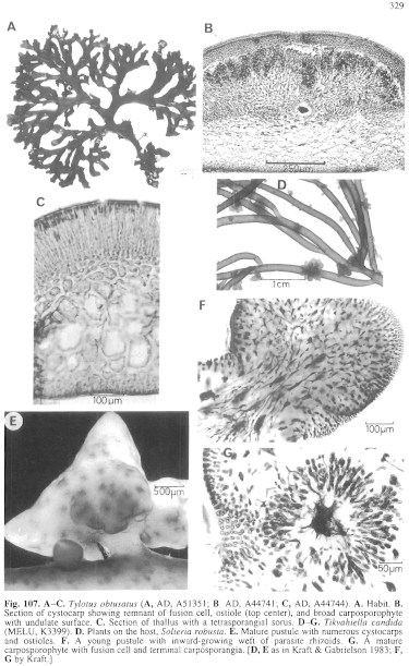
Figure 107 enlarge
Fig. 107. A–C. Tylotus obtusatus (A, AD, A51351; B, AD, A44741; C, AD, A44744). A. Habit. B. Section of cystocarp showing remnant of fusion cell, ostiole (top center), and broad carposporophyte with undulate surface. C. Section of thallus with a tetrasporangial sorus. D–G. Tikvahiella candida (MELU, K3399). D. Plants on the host, Solieria robusta. E. Mature pustule with numerous cystocarps and ostioles. F. A young pustule with inward-growing weft of parasite rhizoids. G. A mature carposporophyte with fusion cell and terminal carposporangia. [D, E as in Kraft & Gabrielson 1983; F, G by Kraft.]
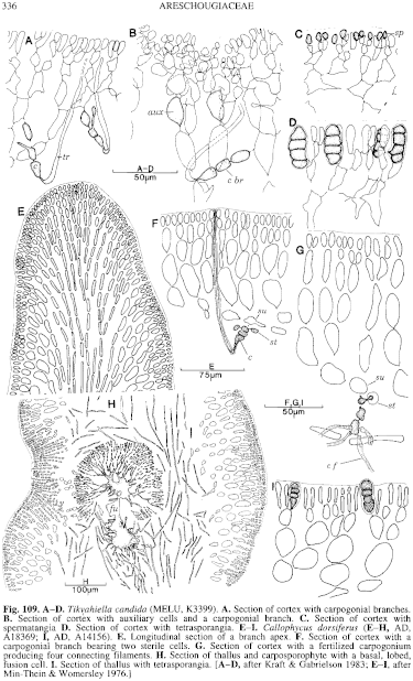
Figure 109 enlarge
Fig. 109. A–D. Tikyahiella candida (MELU, K3399). A. Section of cortex with carpogonial branches. B. Section of cortex with auxiliary cells and a carpogonial branch. C. Section of cortex with spermatangia D. Section of cortex with tetrasporangia. E–I. Callophycus dorsiferus (E–H, AD, A18369; I, AD, A14156). E. Longitudinal section of a branch apex. F. Section of cortex with a carpogonial branch bearing two sterile cells. G. Section of cortex with a fertilized carpogonium producing four connecting filaments. H. Section of thallus and carposporophyte with a basal, lobed, fusion cell. I. Section of thallus with tetrasporangia. [A–D, after Kraft & Gabrielson 1983; E–I, after Min-Thein & Womersley 1976.]

|
Email Contact: State Herbarium of South Australia |

|


