|
|
|
|
|
|||||||||||
|
Electronic Flora of South Australia Species Fact Sheet
Phylum Phaeophyta – Order Dictyotales – Family Scoresbyellaceae
Thallus (Pl. 2 fig. 2; Fig. 92B) medium to dark brown below, light brown near apices, 6–19 cm long, complanate and moderately branched, with a single axis arising from a rhizoidal holdfast 2–5 mm across and 2–5 mm long; epilithic. Fronds branched dichotomously at apices, becoming laterally branched at intervals of (0.2–) 0.5–2 (–3) cm, branches entire, 3–5 mm broad near the truncate to slightly concave apices, mostly 5–10 mm broad throughout the main thallus, decreasing to 1–3 mm broad near their bases. Growth from a single apical cell in an apical depression, lenticular in surface view (Fig. 93A) and rectangular in transverse section (Fig. 93B), segmenting on both lateral sides; daughter cells dividing transversely (Fig. 93A) and then with two walls parallel to the thallus surface, giving three layers (Fig. 93B,C), each row of daughter cells (Fig. 93A) with an apical cell (Fig. 93B) which segments in similar manner and lies on one side of the apical groove, separated from similar cells on the other side by a gelatinous plug. Structure of mature thallus at first with division of the cortical layer giving two layers of fairly densely phaeoplastic cells, the inner layer of which enlarges and loses most of its phaeoplasts, while the central layer becomes distinctly larger (Fig. 93E). Further divisions of the cortical and outer medullary cells produce a 6–8 cell thick medulla (Fig. 93F), with the inner cells progressively larger and arranged more or less in layers but not in regular transverse rows (Fig. 93G). Thallus near apices 60–80 µm and 3 cells thick, slightly thicker near margins (Fig. 93E), mid thallus 150–300 µm and 5–8 cells thick, and near the holdfast biconvex in section, 400–500 µm and 8–12 cells thick (Fig. 93G). Cortical cells (Fig. 93D) in vague lines, becoming irregular, isodiametric to L/B 2, 10–25 (–40) µm across; phaeoplasts discoid, numerous per cell, without pyrenoids. Hair tufts (Fig. 93E) scattered, phaeophycean, external to the cortex; hairs densely clustered, (10–) 12–16 µm in diameter.
Reproduction: Sporangial and oogonial plants unknown. Male plants with scattered, often confluent, sori (Fig. 93H), irregular in shape and mostly 0.5–1.5 mm across, with elongate, sterile paraphyses (Fig. 93I) surrounding the sori. Antheridia (Fig. 93I) densely packed, formed from each cortical cell, elongate, (60–) 80–120 Atm long and 15–25 in diameter.
Type from Egg I., Isles of St Francis, S. Aust., 32–38 m deep (Shepherd, 11.i.1971; ADU, A38067, holotype and isotypes).
Taxonomic notes: Scoresbyella profunda appears to be a rare alga, confined to deep water. It is named after Mr Scoresby A. Shepherd, collector of the type and whose SCUBA collections have contributed greatly to our knowledge of southern Australian deep water algae.
References: The Marine Benthic Flora of Southern Australia Part II
Publication:
Womersley, H.B.S. (14 December, 1987)
The Marine Benthic Flora of Southern Australia
Part II
©Board of the Botanic Gardens and State Herbarium, Government of South Australia
Illustrations in Womersley Part II, 1997: PLATE 2 fig. 2; FIGS 92B, 93.
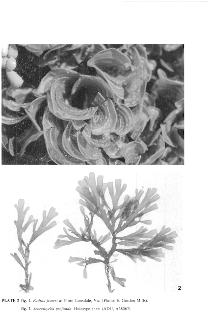
Plate 2 enlarge
PLATE 2 fig. 1. Padina fraseri at Point Lonsdale, Vic. (Photo: E. Gordon-Mills).
fig. 2. Scoresbyella profunda. Holotype sheet (ADU, A38067)
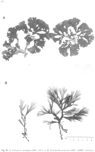
Figure 92 enlarge
Fig. 92. A. Lobophora variegata (ADU, A57111). B. Scoresbyella profunda (ADU, A38067, holotype).
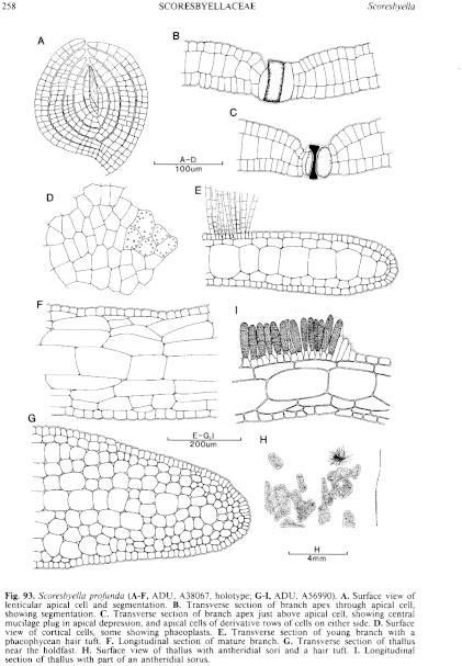
Figure 93 enlarge
Fig. 93. Scoresbyella profunda (A–F, ADU, A38067, holotype; G–I, ADU, A56990). A. Surface view of lenticular apical cell and segmentation. B. Transverse section of branch apex through apical cell, showing segmentation. C. Transverse section of branch apex just above apical cell, showing central mucilage plug in apical depression, and apical cells of derivative rows of cells on either side. D. Surface view of cortical cells, some showing phaeoplasts. E. Transverse section of young branch with a phaeophycean hair tuft. F. Longitudinal section of mature branch. G. Transverse section of thallus near the holdfast. H. Surface view of thallus with antheridial sori and a hair tuft. I. Longitudinal section of thallus with part of an antheridial sorus.

|
Email Contact: State Herbarium of South Australia |

|


