|
|
|
|
|
|||||||||||
|
Electronic Flora of South Australia Species Fact Sheet
Phylum Rhodophyta – Class Florideophyceae – Order Gigartinales – Family Peyssonneliaceae
Selected citations: Boudouresque & Denizot 1975: 58, figs 107–115. Cribb 1983: 41. Denizot 1968: 97, figs 78–82. Schneider & Reading 1987: 185, figs 29–40.
Thallus medium to dark red, (0.5–) 1–2 cm across, orbicular, incompletely adherent to varying extents, with faint concentric growth zones and radial striae, 70–100 µm thick, epilithic or epiphytic. Structure. Basal layer (Fig. 47K) of essentially parallel filaments, 12–18 µm in diameter from below with cells L/D 1.5–2 (–4), 12–16 µm in height and L/D 1–1.5 (–2), often rhomboidal in RVS, with many cells producing unicellular rhizoids 8–12 µm in diameter; hypobasal calcification present. Erect filaments (Fig. 47I) 3–6 (–12) cells long, at (50–) 60–85°, the lowest cell usually more erect but similar (or slightly smaller) in dimensions to the basal layer cell and pit-connected centrally to it, then with two branches (and occasional upper ones), upper cells (8–) 10–12 µm in diameter and L/D 0.5–1, arranged in surface view in distinct rows (Fig. 47J) of square to polygonal cells; internal calcification absent.
Reproduction: Female nemathecia (Fig. 47N) superficial, with paraphyses 8–12 cells long, lower and mid cells 4–6 µm in diameter, L/D 2–5; carpogonial (Fig. 47L) and auxiliary cell branches 3–5 cells long; carposporangia (Fig. 47M–O) in rows of 2–3 on a small gonimoblast cell arising from the fusion cell, more or less isodiametric and 25–35 µm across. Spermatangia unknown (reported on dioecious thalli by Denizot 1968, p. 97).
Tetrasporangia in shallow sori among 5–6-celled paraphyses, 45–60 µm long and 18–30 µm in diameter, cruciately divided.
Type from Gross-Batanga, Cameroon, West Africa, 2–3 m deep; in B?
Distribution: Widely distributed in tropical to warm temperate seas.
In southern Australia, known from the Head of the Great Australian Bight, S. Aust. to D'Entrecasteaux Ch., Tas., in heavily shaded pools to deep water.
Taxonomic notes: The above collections are provisionally placed under P. inamoena. The thallus structure, the usually weak adherence to the substrate, and the regular rows of both the basal layer cells and the upper cells as seen in surface view, all agree with this species and the carposporangial and tetrasporangial nemathecia are very similar to those illustrated by Schneider & Reading (1987, fig. 40).
References:
BOUDOURESQUE, C.-F. & DENIZOT, M. (1975). Révision du genre Peyssonnelia (Rhodophyta) en Méditerranée. Bull. Mus. Hist. Nat. Marseilles 35, 7–92.
CRIBB, A.B. (1983). Marine algae of the southern Great Barrier Reef. Part I. Rhodophyta. (Aust. Coral Reef Soc., Handbook 2: Brisbane.)
DENIZOT, M. (1968). Les Algues Floridées encroûtantes (a l'exclusion des Corallinacées). (Paris.)
PILGER, R. (1911). Die Meeresalgen von Kamerun. Nach der Sammlung von C. Ledermann. In Engler, A., Beitrage zur Flora von Africa xxxix. Bot. Jb. 46, 294–323.
SCHNEIDER, C.W. & READING, R.P. (1987). A revision of the genus Peyssonnelia (Rhodophyta, Cryptonemiales) from North Carolina, including P. atlantica new species. Bull. Mar. Sci. 40, 175–192.
The Marine Benthic Flora of Southern Australia Part IIIA complete list of references.
Publication:
Womersley, H.B.S. (14 January, 1994)
The Marine Benthic Flora of Southern Australia
Rhodophyta. Part IIIA, Bangiophyceae and Florideophyceae (to Gigartinales)
Reproduced with permission from The Marine Benthic Flora of Southern Australia Part IIIA 1994, by H.B.S. Womersley. Australian Biological Resources Study, Canberra. Copyright Commonwealth of Australia.
Illustrations in Womersley Part IIIA, 1994: FIGS 47 1–0.
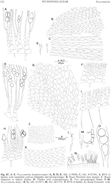
Figure 47 enlarge
Fig. 47. A–E. Peyssonnelia boudouresquei (A, B, D, E, AD, A19608; C, AD, A15184). A. RVS of thallus with separated cortical filaments and tetrasporangia. B. Basal filaments near margin. C. Basal filaments in mature thallus. D. Thallus with carposporangia. E. Two spermatangial heads. F–H. Peyssonnelia dubyi (F, G, AD, A15071; H, AD, A61151). F. RVS of thallus. G. Basal filaments with flabellules. H. RVS of thallus with tetrasporangia. I-0. Peyssonnelia inamoena (AD, A59853). 1. RVS of thallus. J. Surface (upper) pattern of erect filaments. K. Basal filaments. L. Carpogonial branch. M. Young carposporangia from connecting filament. N. Carposporangia in a female nemathecium. 0. Mature carposporangia.
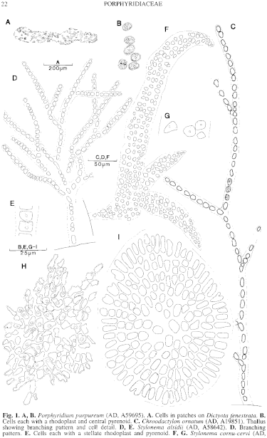
Figure 1 enlarge
Fig. 1. A, B. Porphyridium purpureum (AD, A59695). A. Cells in patches on Dictyota fenestrata. B. Cells each with a rhodoplast and central pyrenoid. C. Chroodactylon ornatum (AD, A19851). Thallus showing branching pattern and cell detail. D, E. Stylonema alsidii (AD, A58642). D. Branching pattern. E. Cells each with a stellate rhodoplast and pyrenoid. F, G. Stylonema cornu-cervi (AD,A58644). F. Thallus (on Caulerpa) showing cell arrangement. G. Cells each with a rhodoplast and pyrenoid. H. Erythrocladia irregularis (on AD, A31991 of Chaetomorpha linum). Thallus showing branching pattern of cells and cell detail. I. Erythrocladia subintegra (AD, A58641). Thallus showing branching pattern of cells and detail in a few.

|
Email Contact: State Herbarium of South Australia |

|


