|
|
|
|
|
|||||||||||
|
Electronic Flora of South Australia Species Fact Sheet
Phylum Phaeophyta – Order Chordariales – Family Chordariaceae
Selected citations: Lindauer et al. 1961: 227, fig. 51(3–5). Nelson & Adams 1983: 80, figs 3,4.
Thallus (Fig. 30B) medium to dark brown, mucoid, densely tomentose, attached by a small discoid holdfast 1–2 mm across, with one to a few long axes with several long laterals and often shorter branchlets, 10–40 cm long and 1–3 mm in diameter, epilithic. Medulla (Fig. 32A) dense, of longitudinal filaments 10–16 (–20) µm in diameter with cells L/B (4–) 6–12 (–15), with only slight production of hyphae except when mature, when the medulla is covered by a weft of hyphal filaments 4–8 µm in diameter. Cortical filaments (Fig. 32A,B) produced directly from the outer medullary filaments near the branch apices, later from the weft of hyphae, of two lengths: shorter determinate filaments 100–250 µm and 10–20 cells long, slightly curved, cells (6–) 8–10 in diameter and L/B 1–2 (–4) above, narrower below; and longer (indeterminate) cortical filaments 1–1.5 mm and usually over 50 cells long, cells 15–20 µm in diameter and L/B 1.5–2 above, becoming narrower (6–8 mm in diameter and L/B 2–4) near their base. Phaeoplasts (Fig. 32C) several per cell, discoid, each with a pyrenoid. Phaeophycean hairs absent.
Reproduction: Sporangia arising directly from the outer medullary filaments or from the hyphal weft; thalli with only unilocular sporangia or with mainly plurilocular but a few unilocular sporangia. Unilocular sporangia (Fig. 32B) borne at base of cortical filaments, scattered or when well developed forming a well-defined layer outside the medulla, clavate to ovoid, (40–) 45–70 (–95) µm long and 15–30 µm in diameter. Plurilocular sporangia (Fig. 32D) terminating branched clusters of slender filaments, the whole forming a dense stratum (100–140 µm broad) at the base of the cortical filaments; sporangia 30–60 pin and 8–25 locules long, 3–4 µm in diameter.
Distribution: New Zealand (see Nelson & Adams 1983, p. 80). East coast of Tasmania.
Taxonomic notes: Type from Paihia, Bay of Islands, New Zealand (Lindauer, 6.xi.1937-"Algae Novae-Zelandicae Exsiccatae" No. 86); in LD, isotypes in other herbaria.
The Tasmanian specimens agree well with the New Zealand species. P. lutea appears to be rare on eastern Tasmanian coasts, but occurred in abundance on wave-washed, sand surrounded, rocks at The Gardens.
Lindauer et al. (1961, p. 228) also credited the South African P. laxa Kylin to New Zealand, but Nelson & Adams (1983, p. 82) place all New Zealand specimens under P. lutea.
However, some New Zealand specimens [e.g. from Motunau Point (Womersley, 15.i.1966; ADU, A29944)] do have considerably broader (22–26 µm) long cortical filaments than given for P. lutea and are more robust plants; they may represent a different entity.
References:
KYLIN, H. (1940). Die Phaeophyceenordnung Chordariales. Acta Univ. lund. N.F. Avd. 2, 36(9), 1–67, Plates 1–8.
LINDAUER, V.W., CHAPMAN, V.J. & AIKEN, M. (1961). The marine algae of New Zealand. II. Phaeophyceae. Nova Hedwigia 3, 129–350, Plates 57–97.
NELSON, W.A. & ADAMS, N.M. (1983). A taxonomic revision of the families Chordariaceae and Chordariopsidaceae (Phaeophyta) in New Zealand. N.Z. J. Bot. 21, 77–92.
The Marine Benthic Flora of Southern Australia Part II complete list of references.
Publication:
Womersley, H.B.S. (14 December, 1987)
The Marine Benthic Flora of Southern Australia
Part II
©Board of the Botanic Gardens and State Herbarium, Government of South Australia
Illustrations in Womersley Part II, 1997: FIGS 30B, 32 A–D.
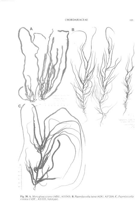
Figure 30 enlarge
Fig. 30. A. Myriogloea sciurus (ADU, A55343). B. Papenfussiella lutea (ADU, A57209). C. Papenfussiella extensa (ADU, A51033, holotype).

Figure 32 enlarge
Fig. 32. A–D. Papenfussiella lutea (ADU, A57209). A. Longitudinal section (partly diagrammatic) showing medullary filaments, short (determinate) and long (indeterminate) cortical filaments, stratum of plurilocular sporangia, and unilocular sporangia. B. Medullary and hyphal filaments with long and short cortical filaments and young unilocular sporangia. C. Cells of a long (indeterminate) cortical filament with phaeoplasts and pyrenoids. D. Medullary hyphal filament with branch systems bearing plurilocular sporangia. E–G. Papenfussiella extensa (ADU, A51033, holotype). E. Transverse section showing medullary cells, short (determinate) and long (indeterminate) cortical filaments and unilocular sporangia. F. Longitudinal section, as for E. G. Cells of a long (indeterminate) cortical filament with phaeoplasts and pyrenoids. H–J. Suringariella harveyana (H,I, ADU, A24576; J, ADU, A30676). H. Outer medulla and cortex with simple or branched determinate cortical filaments, a phaeophycean hair, and unilocular sporangia. I. Cells of a determinate cortical filament with phaeoplasts (with pyrenoids) and physodes. J. Determinate cortical filaments with plurilocular sporangia of various ages.

|
Email Contact: State Herbarium of South Australia |

|


