|
|
|
|
|
|||||||||||
|
Electronic Flora of South Australia Species Fact Sheet
Phylum Phaeophyta – Order Dictyosiphonales – Family Myriotrichiaceae
Selected citations: Pedersen 1978: 281, figs 1–34. Rosenvinge & Lund 1947: 50, figs 17, 18. Taylor 1957: 161, p1. 10 figs 4–7.
Synonyms
M. filiformis Harvey 1848: pl. 156. Kylin 1947: 70, fig. 58A,B.
M. repens (Hauck) Karsakoff. Kuckuck 1899: 55, figs 1–4, pl. 3. Kylin 1947: 71, fig. 58C–G.
Thallus (Fig. I IOC) with prostrate, branched, largely uniseriate filaments in the outer tissue of the host, cells usually swollen, (8–) 10–15 µ in diameter and L/B 1–1.5 (–2), producing numerous erect, simple, axes. Growth apical or trichothallic in young axes, becoming intercalary. Structure. Axes 0.2–1.0 mm high, uniseriate below and 10–12 µm in diameter, cells L/B 2–3, becoming multiseriate above (2–3 cells thick) in at least some segments by longitudinal and oblique divisions, 15–20 µm in diameter with cells L/B 0.4–1.0, tapering slightly to the apices of the erect axes; cells with several phaeoplasts, each with a pyrenoid. Phaeophycean hairs numerous, terminal and lateral, usually single, scattered, 5–8 1.1m in diameter.
Reproduction: Plurilocular sporangia (Fig. 1 10C) formed laterally and radially from upper cells of erect filaments, clustered, uniseriate with occasional oblique divisions, simple or basally branched, tapering, 15–25 µm and 4–6 locules long, 4–8 µm in diameter. Unilocular sporangia (Fig. 110C) sessile, single, paired or clustered on the subapical cell of erect axes just below the terminal hair, subspherical to ovoid, 20–30 µm long and 15–25 µm in diameter.
Type from Torquay, England, on Scytosiphon (Griffiths, Aug. 1833); in Herb. Harvey, TCD?
Distribution: Widely distributed in cool temperate European and eastern N. American coasts and the Mediterranean, epiphytic on various algae, especially Chordariales. Recorded from Florida by Earle (1969, p. 196, as M. repens).
Taxonomic notes: The southern Australian collections correspond to M. repens in their small size, and to M. filiformis in that the erect axes are mostly uniseriate, with longitudinal divisions occasional in the basal filaments and only in the upper parts of the erect axes which are becoming fertile. Loiseaux (1969, p. 11) found that zooids from plurilocular sporangia of Hecatonema maculans gave rise to thalli characteristic of Myriotrichia when cultured at 12°C, and Pedersen (1978) has shown that M. filiformis and M. repens are growth forms of M. clavaeformis, usually produced under sub-optimal conditions of salinity and light. Pedersen also considers that the prostrate system of M. clavaeformis is very similar to Streblonema sphaericum, which is usually placed in the Ectocarpales.
The two southern Australian collections may be relatively poorly developed plants, and may well be connected with plants provisionally placed in Hecatonema. M. adriatica Hauck was recorded from southern New Zealand by Lindauer et al. (1961, p. 264).
References:
EARLE, S.A. (1969). Phaeophyta of the Eastern Gulf of Mexico. Phycologia 7, 71–254.
HARVEY, W.H. (1834). Algological Illustrations. I. Remarks on some British algae, and descriptions of new species recently added to our flora. .1. Bot. (Hooker) 1, 296–305, Plates 188, 189.
HARVEY, W.H. (1847). Phycologia Britannica. Plates 73–144. (Reeve: London.)
HARVEY, W.H. (1848). Phycologia Britannica. Plates 145–216. (Reeve: London.)
KUCKUCK, P. (1899). Beiträge zur Kenntnis der Meeresalgen. 9. Ober den Generationswechsel von Cutleria multifida (Engl. Bot.) Grev. Wiss. Meeresunters. Abt. Helgol., N.F. 3, 95–117.
KYLIN, H. (1947). Die Phaeophyceen der Schwedischen Westkiiste. Acta Univ. lund. N.F. Avd. 2, 43(4), 1–99, Plates 1–18.
LINDAUER, V.W., CHAPMAN, V.J. & AIKEN, M. (1961). The marine algae of New Zealand. II. Phaeophyceae. Nova Hedwigia 3, 129–350, Plates 57–97.
LOISEAUX, S. (1969). Sur une espèce de Myriotrichia obtenue en culture à partir de zoides d'Hecatonema maculans Sauv. Phycologia 8, 11–15.
PEDERSEN, P.M. (1978). Culture studies in the pleomorphic brown alga Myriotrichia clavaeformis (Dictyosiphonales, Myriotrichiaceae). Norw. J. Bot. 25, 281–291.
ROSENVINGE, L.K. & LUND, S. (1947). The marine algae of Denmark. Vol. II. Phaeophyceae. III. Encoeliaceae, Myriotrichiaceae, Giraudiaceae, Striariaceae, Dictyosiphonaceae, Chordaceae and Laminariaceae. K. Dan. Vidensk. Selsk. Biol. Skr. 4(5), 1–99.
TAYLOR, W.R. (1957). Marine algae of the Northeastern coast of North America. Rev. Edn. (Univ. Mich. Press: Ann Arbor.)
The Marine Benthic Flora of Southern Australia Part II complete list of references.
Publication:
Womersley, H.B.S. (14 December, 1987)
The Marine Benthic Flora of Southern Australia
Part II
©Board of the Botanic Gardens and State Herbarium, Government of South Australia
Illustration in Womersley Part II, 1997: FIG. 110C.
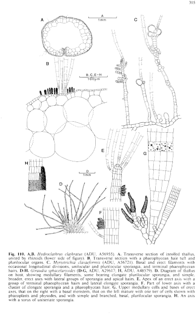
Figure 110 enlarge
Fig. 110. A,B. Hydroclathrus clathratus (ADU, A56955). A. Transverse section of inrolled thallus, united by rhizoids (lower side of figure). B. Transverse section with a phaeophycean hair tuft and plurilocular organs. C. Myriotrichia clavaefortnis (ADU, A56721). Basal and erect filaments with occasional longitudinal divisions, unilocular and plurilocular sporangia, and terminal phaeophycean hairs. D–H. Giraudia sphacelarioides (D–G, ADU, A29617; H, ADU, A48579). D. Diagram of thallus on host, showing medullary filaments, some bearing elongate plurilocular sporangia, and simple, broader, erect axes with lateral groups of sporangia and apical hairs. E. Apex of an erect axis with a group of terminal phaeophycean hairs and lateral elongate sporangia. F. Part of lower axis with a cluster of elongate sporangia and a phaeophycean hair. G. Upper medullary cells and bases of erect axes, that on the right with a basal meristem, that on the left mature with one tier of cells shown with phaeoplasts and physodes, and with simple and branched, basal, plurilocular sporangia. H. An axis with a sorus of uniseriate sporangia.

|
Email Contact: State Herbarium of South Australia |

|


