|
|
|
|
|
|||||||||||
|
Electronic Flora of South Australia Species Fact Sheet
Phylum Rhodophyta – Family Rhodomelaceae – Tribe Lophothalieae
Selected citations: Shepherd & Womersley 1981: 367.
Thallus (Fig. 104A) light to dark red-brown, fading to yellow-brown, mucilaginous, 5–15 (–20) cm high, with 1 to several corticated erect axes bearing few to many, irregularly radial, lateral branches for 1–2 orders, axes terete, 2–4 mm in diameter (including trichoblasts) densely clothed with branched rhodoplastic trichoblasts except close to the thallus base. Attachment by a mass of tissue within the host conceptacles; epiphytic on Cystophora siliquosa. Structure monopodial (Fig. 105A), apical cells dome-shaped, trichoblasts arising on a 1/5 spiral close to the apices with 5 pericentral cells cut off in alternating sequence from 6–8 segments below apices. Pericentral cells cut off from 9–12 segments below apices, divide transversely, the upper cell forming an adventitious filament. Young branches 35–60 µm in diameter with segments L/D 0.6–1, corticated branches 0.5–2 mm in diameter. Trichoblasts (1–) 2–3 mm long, on each segment in a Is spiral, basally branched 1–2 times with long unbranched ends, developing into monopodial axes of short shoots bearing once furcate secondary trichoblasts (Fig. 105C), with 2–5 basal cells 15–20 µm in diameter and L/D 1.5–2, mid and upper cells 20–40 (–55) µm in diameter and L/D 4–10 (–15), tapering slightly above. Cortication commencing 18–20 segments from apices, becoming heavy with the pericentral cells lost in sectional view (Fig. 105B) and most outer cortical cells bearing simple adventitious filaments with 2–5 small basal cells, upper cells of similar dimensions to trichoblasts. Lateral branches arising by further development of short shoots. Cells uni- or multinucleate; rhodoplasts discoid.
Reproduction: Gametophytes dioecious. Procarps (Fig. 105D) occur on segment 4 or 5 of adventitious filaments, with the fifth pericentral cell bearing a sterile group initial and a 4-celled carpogonial branch. Carposporophytes with a basal fusion cell and branched gonimoblast with clavate terminal carposporangia 15–25 µm in diameter. Cystocarps (Fig. 104B, C) urceolate, sessile or short stalked, 450–700 µm in diameter, with a slight neck; pericarp ostiolate, with about 12 erect filaments, each cell cutting off 2 isodiametric outer cells and with a cortex 1–2 cells thick. Spermatangial organs (Fig. 104D) developed from branches of trichoblasts or the ends of short adventitious filaments, 90–220 µm long and 35–55 µm in diameter, with a 4–5 celled monosiphonous stalk and 1–2 sterile terminal cells; three pericentral cells (Fig. 105E) divide to form a layer of initials which cut off an outer layer of spermatang i a.
Tetrasporangial stichidia (Fig. 104E) borne on the simple adventitious filaments, 180–350 µm long and 85–110 µm in diameter, with 4 pericentral cells (Fig. 105G), the second produced forming a straight row (Fig. 105F) of tetrasporangia each with 2 pre-sporangial and 1 post-sporangial cover cells (Fig. 105G); tetrasporangia 50–80 µm in diameter.
Type from Pennington Bay, Kangaroo I., S. Aust., on Cystophora siliquosa, sublittoral fringe (Parsons, 17.xi.1967); holotype in AD, A31891-"Marine Algae of southern Australia" No. 179a). Isotypes distributed under this number.
Selected specimens: Elliston, S. Aust., on Cystophora siliquosa, 5 m deep (Shepherd, 28.x.1972; AD, A42838). Sleaford Bay, S. Aust., on C. siliquosa, sublittoral fringe (Womersley, 16.ii.1959; AD, A22490). Wanna, S. Aust., on C. siliquosa, upper sublittoral pools (Womersley, 19.ii.1959; AD, A22435). Vivonne Bay, Kangaroo I., S. Aust., drift (Womersley, 30.i.1956; AD, A20171). Pennington Bay, Kangaroo I., S. Aust., on C. siliquosa, sublittoral fringe (Womersley, 19.i.1965; AD, A28927-"Marine Algae of southern Australia" No. 179b) and on C. intermedia, sublittoral fringe (Womersley, 9.x.1997; AD, A67286). Bridgewater Bay, Vic., on C. siliquosa, upper sublittoral (Beauglehole, 26.xii.1950; AD, A15686).
Distribution: Elliston, S. Aust., to Bridgewater Bay, Victoria.
Taxonomic notes: H. tomentosa differs from H. urceolata in habit, in having numerous adventitious filaments produced from divided pericentral cells and cortical cells, and in growing mainly on Cystophora siliquosa whereas the latter occurs on other species of Cystophora.
References:
PARSONS, M.J. (1975). Morphology and taxonomy of the Dasyaceae and Lophothalieae (Rhodomelaceae) of the Rhodophyta. Aust. J. Bot. 23(4), 549–713.
SHEPHERD, S.A. & WOMERSLEY, H.B.S. (1981). The algal and seagrass ecology of Waterloo Bay, South Australia. Aquat. Bot. 11, 305–371.
The Marine Benthic Flora of Southern Australia Part IIID complete list of references.
Publication:
Womersley, H.B.S. (24 February, 2003)
The Marine Benthic Flora of Southern Australia
Rhodophyta. Part IIID. Ceramiales – Delesseriaceae, Sarcomeniaceae, Rhodomelaceae
Reproduced with permission from The Marine Benthic Flora of Southern Australia Part IIID 2003, by H.B.S. Womersley. Australian Biological Resources Study, Canberra. Copyright Commonwealth of Australia.
Illustrations in Womersley Part IIIA, 2003: FIGS 104, 105.
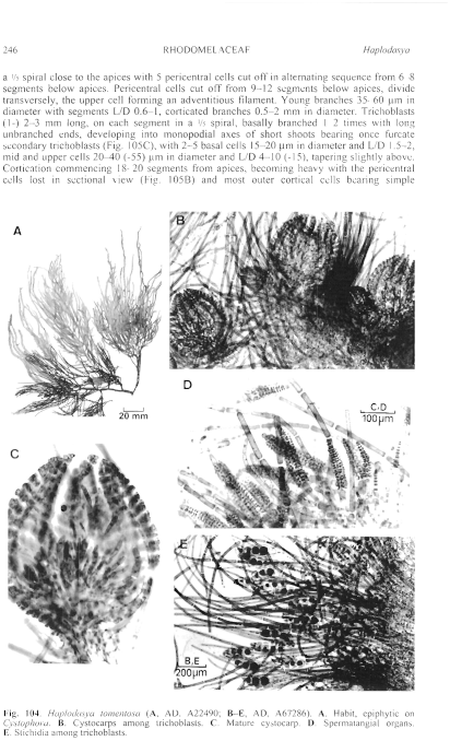
Figure 104 enlarge
Fig. 104. Haplodasya tomentosa (A, AD, A22490; B–E, AD, A67286). A. Habit, epiphytic on Cystophora. B. Cystocarps among trichoblasts. C. Mature cystocarp. D. Spermatangial organs. E. Stichidia among trichoblasts.
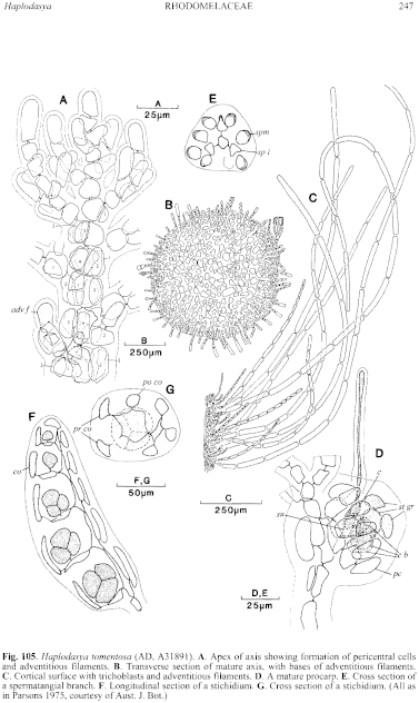
Figure 105 enlarge
Fig. 105. Haplodasya tomentosa (AD, A31891). A. Apex of axis showing formation of pericentral cells and adventitious filaments. B. Transverse section of mature axis, with bases of adventitious filaments. C. Cortical surface with trichoblasts and adventitious filaments. D. A mature procarp. E. Cross section of a spermatangial branch. F. Longitudinal section of a stichidium. G. Cross section of a stichidium. (All as in Parsons 1975, courtesy of Aust. J. Bot.)

|
Email Contact: State Herbarium of South Australia |

|


