|
|
|
|
|
|||||||||||
|
Electronic Flora of South Australia Species Fact Sheet
Phylum Rhodophyta – Class Florideophyceae – Order Gelidiales – Family Gelidiaceae
Selected citations: Boudouresque 1972: 1, figs 1–10. Maggs & Guiry 1987: 429. Norris 1992b: 35, fig. 21.
Synonym
G. stichidiospora Dawson 1953: 84, pl. 12 figs 4,5.
Thallus (Fig. 34K) medium red-brown, growing within a turf of other algae, with prostrate stolons producing simple erect branches 1–2 (–3) mm high. Stolons terete, 50–70 µm in diameter, tapering to the apical cell, attached by a fringe of rhizoids (single from each cell) along much of their length, clumped below erect branches; cortical cells isodiametric to transversely slightly elongate, 8–10 µm across. Erect branches subterete to slightly compressed, closely adjacent to separated on the stolons, simple (rarely once branched), 40–75 µm broad, tapering to a conspicuous apical cell. Holdfast of clumped rhizoids; epilithic or on limpets or abalone. Structure uniaxial, with a fairly prominent axial filament (Fig. 34L, M) cutting off 2–4 periaxial cells which cut off further cells laterally resulting in a compressed branch (Fig. 34L), and forming a medulla 2–3 cells broad of elongate cells 4–6 µm in diameter and a cortex 1–2 cells thick, the outer cells (Fig. 34M) more or less in rows in surface view, quadrangular, lengthening to L/B 1–1.5 (–2) and 5–8 µm across, each with several small rhodoplasts; rhizines absent.
Reproduction: Sexual plants unknown.
Tetrasporangial stichidia (Fig. 34M) terminating erect branches, 70–90 µm in diameter and (150–) 200–600 [tin long, terete; tetrasporangia formed in regular sequence from the apex, 4 per segment (Fig. 34N), cut off from each of 4 periaxial cells and with two layers of cortical cells, the outer layer of small cells with gaps through which the tetraspores escape; tetrasporangia subspherical, 25–35 µm in diameter, irregularly decussately divided.
Type from Cape Caliacra, Black Sea, Romania; in ?
Distribution: Apart from the type, known from the Mediterranean (Verlaque 1990a, p. 82); from Isla Cedros, Baja California (Dawson) on Haliotis; and from Natal, South Africa (Norris).
In southern Australia, from Topgallant I. to Vivonne Bay, Kangaroo I., S. Aust.
Taxonomic notes: The Australian collections of this minute turf species agree very well with the description by Boudouresque, and extend further its distribution; it may well be widely distributed but overlooked in most turf collections. The only apparently related species listed by Maggs & Guiry (1987, p. 429) is G. adnata Dawson (1954, p. 422, fig. 33f) from Vietnam, which has tetrasporangia in tiers of 4, but in stichidia borne directly on the stolon; Dawson's figure lacks detail. Norris (1992b, p. 35) considers G. adnata a synonym of G. antipai. Cribb (1983, p.31) placed both G. adnata and G. stichidiospora under G. pannosa, but the latter shows 4–5 sporangia in surface view and hence has whorls of 8–10 sporangia (Feldmann & Hamel 1936, fig. 12B, as G. tenuissima).
References:
BOUDOURESQUE, C.-F. (1972). Végétation marine de l'île de Port-Cros (Parc National) ix. - Sur Gelidiella antipai Marie Celan (Gélidiales). Bull. Soc. phycol. Fr. 17, 1–8.
CELAN, M. (1938). Notes sur la flore algologique du littoral Roumain de la Mer Noire. IV. Deux Rhodophycées nouvelles pour la flore de la Mer Noire: Gelidiella antipae et Phyllophora brodiaei (Turn.)J.Ag. Bull. Sect. sci. Acad. roum. 19, 76–79.
CRIBB, A.B. (1983). Marine algae of the southern Great Barrier Reef. Part I. Rhodophyta. (Aust. Coral Reef Soc., Handbook 2: Brisbane.)
DAWSON, E.Y. (1953). Marine red algae of Pacific Mexico. Part I. Bangiales to Corallinaceae subf. Corallinoideae. Allan Hancock Pacif. Exped. 17, 1–239.
DAWSON, E.Y. (1954). Marine plants in the vicinity of the Institut Océanographique de Nha Trang, Viêt Nam. Pacific Sci. 8, 372–481.
FELDMANN, J. & HAMEL, G. (1936). Floridées de France VII. Gélidiales. Rev. Alg. 9, 85–140, Plates 2–6.
MAGGS, C.A. & GUIRY, M.D. (1987). Gelidiella calcicola sp. nov. (Rhodophyta) from the British Isles and northern France. Br. phycol. J. 22, 417–434.
NORRIS, R.E. (1992b). The marine red algae of Natal, South Africa: Order Gélidiales (Rhodophyta). Mem. Bot. Survey South Africa No. 61.
VERLAQUE, M. (1990a). Végétation marine de la Corse (Méditerranée). VIII. Documents pour la flore des algues. Vie Milieu 40, 79–92.
The Marine Benthic Flora of Southern Australia Part IIIA complete list of references.
Publication:
Womersley, H.B.S. (14 January, 1994)
The Marine Benthic Flora of Southern Australia
Rhodophyta. Part IIIA, Bangiophyceae and Florideophyceae (to Gigartinales)
Reproduced with permission from The Marine Benthic Flora of Southern Australia Part IIIA 1994, by H.B.S. Womersley. Australian Biological Resources Study, Canberra. Copyright Commonwealth of Australia.
Illustration in Womersley Part IIIA, 1994: FIG. 34 K–N.
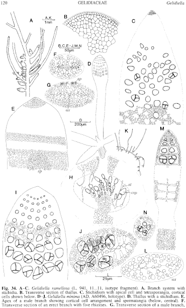
Figure 34 enlarge
Fig. 34. A–C. Gelidiella ramellosa (L, 941, 11...11, isotype fragment). A. Branch system with stichidia. 13. Transverse section of thallus. C. Stichidium with apical cell and tetrasporangia, cortical cells shown below. D–J. Gelidiella minima (AD, A60496, holotype). D. Thallus with a stichidium. E. Apex of a male branch showing cortical cell arrangement and spermatangia (below, central). F. Transverse section of an erect branch with five rhizines. G. Transverse section of a male branch, showing axial cell, secondary transverse row and cortical cells; spermatangia on upper surface. H. Section of a cystocarp, showing carposporangia, elongate arachnoid cells and ostiole. I. Surface view of a stichidium with tetrasporangia. J. Cross section of a stichidium. K–N. Gelidiella antipai (AD, A59079). K. Thallus with stolon and erect branches. L. Cross section of branch showing axial cell, secondary row, and cortical cells. M. A stichidium (segments omitted at dashes) showing tetrasporangial whorls and cortical cells (below). N. Cross section of stichidium with a whorl of four tetrasporangia. [D, E, G–J also used in Guiry & Womersley 1992]

|
Email Contact: State Herbarium of South Australia |

|


