|
|
|
|
|
|||||||||||
|
Electronic Flora of South Australia Species Fact Sheet
Phylum Rhodophyta – Family Rhodomelaceae – Tribe Lophothalieae
Selected citations: De Toni 1903: 1022. Lucas 1909: 43; 1929b: 51. Lucas & Perrin 1947: 287, fig. 132. May 1965: 378. Parsons 1975: 665, figs 32, 33, 46B. Shepherd & Womersley 1981: 367.
Synonym
Lophothalia lanuginosa J. Agardh 1890: 64, pl. 2 fig. 3. Reinbold 1898: 50. Schmitz 1893: 220.
Thallus (Fig. 114C) dark red-brown, (10–) 20–70 cm high, with a single main axis bearing denuded, corticated, lower branches 1–2 mm in diameter, with clustered upper branches 10–40 cm long, branched for 2–3 orders, densely and evenly clothed (Fig. 114D) with rhodoplastic trichoblasts and monosiphonous filaments 1–3.5 mm long. Holdfast at first discoid, soon becoming fibrous and 1–2 cm across; epilithic. Structure monopodial, with trichoblasts arising 1–3 axial cells apart and 4 pericentral cells cut off in alternating order 20–25 axial cells below apices. Adventitious monosiphonous filaments arise (Fig. 116A) from pericentral and cortical cells and develop a short determinate axis of 6–12 isodiametric cells bearing trichoblasts (Fig. 116B) spirally, with short basal cells, mid and upper cells 15–25 µm in diameter and L/D 3–7, terminal cells shorter with rounded ends; these short shoots with trichoblasts densely and evenly clothe the branches, some becoming indeterminate and developing into long lateral branches. Cortication starting about 40 segments from apices and becoming heavy on lower branches, with the 4 pericentral cells (Fig. 114E) remaining clear in transverse section, surrounded by larger inner and small outer cortical cells. Cells multinucleate; rhodoplasts discoid, becoming chained or ribbon like.
Reproduction: Gametophytes dioecious. Procarps (Fig. 116C) developed 2–3 cells from base of a trichoblast, with 5 pericentral cells, the fifth producing a sterile group of 4 cells and a 4-celled carpogonial branch. Carposporophyte with a basal fusion cell and erect, branched, gonimoblast filaments with clavate terminal carposporangia 25–40 µm in diameter, replaced from below. Cystocarps (Fig. 116D) subglobose, 1–1.5 mm in diameter, with a stalk 0.5–2 mm long and no neck; pericarp ostiolate, 5–7 cells thick with small outer cortical cells. Spermatangial organs (Fig. 116E) developed on trichoblasts, with long, sterile basal and terminal filaments, 200–300 µm long and 30–50 µm in diameter, each segment with 3 pericentral cells (Fig. 116F), dividing to 5, producing initials and an outer layer of spermatangia.
Tetrasporangial stichidia (Fig. 116G) developed from axes of short shoots with few trichoblasts, on a monosiphonous base, 300–600 (–1000) µm long and 60–160 µm in diameter, with 4 pericentral cells producing decussate pairs of tetrasporangia 45–75 µm in diameter, each with 2 pre-sporangial and 1 post-sporangial cover cells.
Lectotype from Encounter Bay, S. Aust. (Hussey); in Herb. Agardh, LD, 42150.
Selected specimens: Esperance, W. Aust., drift (Burbidge, 30.iii.1937; UWA, A844). Golden I., Avoid Bay, S. Aust., 22 m deep (Hone, 28.ix.1989; AD, A59810). Elliston, S. Aust., drift (Woelkerling, 28.xii.1976; AD, A47870). Port Elliot, S. Aust., drift (Parsons, 15.i.1968; AD, A32205-"Marine Algae of southern Australia" No. 129b, and AD, A32206). Seal Bay, Kangaroo I, S. Aust., drift (Womersley, 21.i.1965; AD, A28676-"Marine Algae of southern Australia" No. 129a). Robe, S. Aust., drift (Womersley, 26.i.1967; AD, A31213). Stinky Bay, Nora Creina, S. Aust., drift (Womersley, 16.v.1982; AD, A55541). 2 km N of Blackfellows Caves, S. Aust., drift (Womersley, 24.xi.1992; AD, A62208). Lady Bay, Vic., (March 1857; MEL 106625). Mussleroe Bay, NE Tas. (G. & F. Perrin, Mar. 1937; HO, 47650, 47651).
Distribution: Esperance, W. Aust., to Lady Bay (Warrnambool), Vic. and NE Tasmania.
Taxonomic notes: D. lanuginosa was described in detail by Parsons (1975, p. 665). It differs from D. bolbochaete in having a denser and more even cover of trichoblasts and short shoots over the branches.
References:
AGARDH, J.G. (1890). Till algernes systematik. Acta Univ. lund. 26(3), 1–125, Plates 1–3.
DE TONI, G.B. (1903). Sylloge Algarum omnium hucusque Cognitarum. Vol. 4. Florideae. Sect. 3. pp. 775–1521 + 1523–1525. (Padua.)
FALKENBERG, P. (1901). Die Rhodomelaceen des Golfes von Neapel und der angrenzenden Meeres-abschnitte. Fauna und Flora des Golfes von Neapel. Monogr. 26. (Friedländer: Berlin.)
LUCAS, A.H.S. & PERRIN, F. (1947). The Seaweeds of South Australia. Part 2. The Red Seaweeds. (Govt Printer: Adelaide.)
LUCAS, A.H.S. (1909). Revised list of the Fucoideae and Florideae of Australia. Proc. Linn. Soc. N.S.W. 34, 9–60.
LUCAS, A.H.S. (1929b). A census of the marine algae of South Australia. Trans. R. Soc. S. Aust. 53, 45–53.
MAY, V. (1965). A census and key to the species of Rhodophyceae (red algae) recorded from Australia. Contr. N.S. W. Natl Herb. 3, 349–429.
PARSONS, M.J. (1975). Morphology and taxonomy of the Dasyaceae and Lophothalieae (Rhodomelaceae) of the Rhodophyta. Aust. J. Bot. 23(4), 549–713.
REINBOLD, T. (1898). Die Algen der Lacepede und Guichen Bay (Süd Australien) und deren näherer Umgebung, gesammelt von Dr. A. Engelhart-Kingston. II. Nuova Notarisia 9, 33–54.
SCHMITZ, F. (1893). Die gattung Lophothalia, J. Ag. Ber. Deutsch. Bot. Ges. 11, 212–232.
SHEPHERD, S.A. & WOMERSLEY, H.B.S. (1981). The algal and seagrass ecology of Waterloo Bay, South Australia. Aquat. Bot. 11, 305–371.
The Marine Benthic Flora of Southern Australia Part IIID complete list of references.
Publication:
Womersley, H.B.S. (24 February, 2003)
The Marine Benthic Flora of Southern Australia
Rhodophyta. Part IIID. Ceramiales – Delesseriaceae, Sarcomeniaceae, Rhodomelaceae
Reproduced with permission from The Marine Benthic Flora of Southern Australia Part IIID 2003, by H.B.S. Womersley. Australian Biological Resources Study, Canberra. Copyright Commonwealth of Australia.
Illustrations in Womersley Part IIIA, 2003: FIGS 114 C–E, 116.
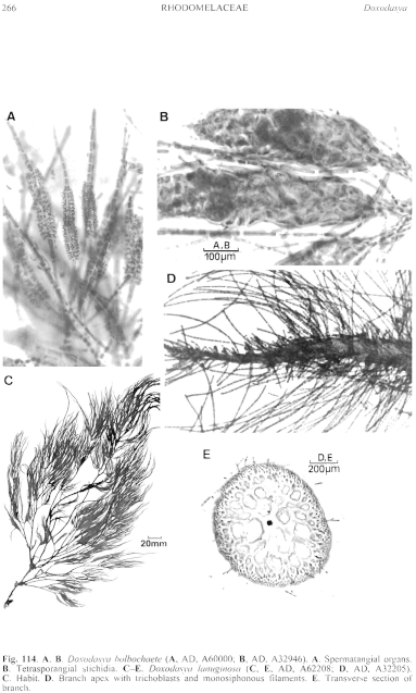
Figure 114 enlarge
Fig. 114. A, B. Doxodasya bolbochaete (A, AD, A60000; B, AD, A32946). A. Spermatangial organs. B. Tetrasporangial stichidia. C–E. Doxodasya lanuginosa (C, E, AD, A62208; D, AD, A32205). C. Habit. D. Branch apex with trichoblasts and monosiphonous filaments. E. Transverse section of branch.
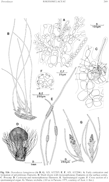
Figure 116 enlarge
Fig. 116. Doxodasya lanuginosa (A–D, G, AD, A32205; E, F, AD, A32206). A. Early cortication and formation of adventitious filaments. B. Short shoots with monosiphonous filaments on the surface cortex. C. Procarp. D. Cystocarp and monosiphonous filaments. E. Spermatangial organs. F. Cross section of a spermatangial organ. G. Mature stichidia. (All as in Parsons 1975, courtesy of Aust. J. Bot.)

|
Email Contact: State Herbarium of South Australia |

|


