|
|
|
|
|
|||||||||||
|
Electronic Flora of South Australia Species Fact Sheet
Phylum Phaeophyta – Order Chordariales – Family Leathesiaceae
Thallus (Figs 27C, 28E) light brown, subglobose to irregularly lobed, mucoid and soft, 1–10 mm across, epiphytic on Cystophora brownii and C. botryocystis. Basal filaments diffuse, irregularly branched, with small, isodiametric cells adnate to but not penetrating the host meristoderm. Medulla (Fig. 28E) extensive, 1–3 (–7) mm high, of branched, cylindrical filaments widely separated in the mucilage, with di- to quadri-chotomous branching every (1–) 2–5 cells, cells (8–) 10–20 (–25) µm in diameter and L/B (2–) 3–7 (–9), sometimes slightly swollen. Deter>minate cortical filaments (Fig. 28F,G) continuing from medullary filaments or in basally branched clusters, densely aggregated and forming a stratum 300–500 ALM and 20–40 cells high, simple, curved above with cells inflated on their upper side, lower cells cylindrical, 6–8 µm in diameter and L/B 2–3 (–4), upper cells 8–10 µm in diameter and L/B (0.5–) 1–1.5 (–2). Phaeoplasts (Fig. 28G) few (2–3) per cell, discoid, each with a pyrenoid. Phaeophycean hairs scattered, arising from upper medullary cells, 10–15 µm in diameter.
Reproduction: Plurilocular sporangia (Fig. 28F,G) borne on the upper cells of the determinate cortical filaments, uniseriate or biseriate, elongate-clavate, 30–60 µm and 6–9 (–16) locules long, 6–8 µm in diameter. Unilocular sporangia unknown.
Type from Point Westall, S. Aust., on Cystophora brownii in lower eulittoral pools (Skinner, 30.xi.1977); holotype in ADU, A48886.
Distribution: Known from the type and Partney I., Sir Joseph Banks Group, S. Aust., on C. botryocystis, 10–12 m deep (Baldock, 13.xii.1977; ADU, A48925). Reevesby I., Sir Joseph Banks Group, S. Aust., on C. brownii, 0–3.5 m deep (Baldock, 13.xii.1977; ADU, A48924). Aldinga, S. Aust., on C. brownii, sublittoral (Skinner, 26.x.1977; ADU, A48630).
Taxonomic notes: Corynophlaea cristata is named from the distinctive cristate appearance of the plurilocular sporangia on the curved parts of the cortical filaments. No other species of the genus has such plurilocular sporangia, and the thalli are usually much larger than in C. cystophorae. They also have almost cylindrical, much less closely adjacent, medullary filaments compared to those of the latter species.
References: The Marine Benthic Flora of Southern Australia Part II
Publication:
Womersley, H.B.S. (14 December, 1987)
The Marine Benthic Flora of Southern Australia
Part II
©Board of the Botanic Gardens and State Herbarium, Government of South Australia
Illustration in Womersley Part II, 1997: FIG. 28 E–G.
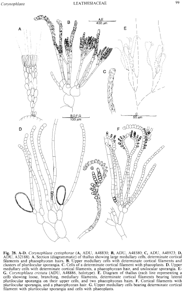
Figure 28 enlarge
Fig. 28. A–D. Corynophlaea cystophorae (A, ADU, A48830; B, ADU, A48580; C, ADU, A48923; D, ADU, A32188). A. Section (diagrammatic) of thallus showing large medullary cells, determinate cortical filaments and phaeophycean hairs. B. Upper medullary cells with determinate cortical filaments and clusters of plurilocular sporangia. C. Cells of a determinate cortical filament with phaeoplasts. D. Upper medullary cells with determinate cortical filaments, a phaeophycean hair, and unilocular sporangia. E–G. Corynophlaea cristata (ADU, A48886, holotype). E. Diagram of thallus (each line representing a cell) showing loose, branching, medullary filaments, determinate cortical filaments bearing lateral plurilocular sporangia on their upper cells, and two phaeophycean hairs. F. Cortical filaments with plurilocular sporangia, and a phaeophycean hair. G. Upper medullary cells bearing determinate cortical filament with plurilocular sporangia and cells with phaeoplasts.

|
Email Contact: State Herbarium of South Australia |

|


