|
|
|
|
|
|||||||||||
|
Electronic Flora of South Australia Species Fact Sheet
Phylum Rhodophyta – Order Ceramiales – Family Ceramiaceae – Tribe Antithamnieae
Selected citations: Kendrick et al. 1988: 204. Norris 1987: 19, figs 1–5? Silva et al. 1996: 377. Stegenga 1986: 28, pl. 4. Stegenga et al. 1997: 381.
Thallus (Figs 47D, 48A) with prostrate axes bearing numerous erect lateral branches up to 4 mm long, with opposite, decussate, whorl-branchlets (pinnae) from each axial cell, curving upwards towards the branch apex. Attachment by rhizoids from the basal cells of pinnae with digitate haptera; epiphytic on geniculate coralline algae. Structure. Apical cells 4–5 µm in diameter and L/D 0.7–1.2, surrounded by young whorl-branchlets and enlarging to 60–90 µm (including a thick sheath) in diameter and L/D 0.7–2 in mature axial cells. Pinnae (Fig. 47E) 200–450 (–580) µm and 10–15 cells long, basal rachis cells 25–32 µm in diameter and L/D 1–1.5, tapering to terminal cells 6–9 µm in diameter and L/D 1–1.5, each of the lower 2 (1–3) cells with opposite pinnules and upper cells with alternate pinnules, pinnules 10–22 µm in diameter, cells L/D 1–1.5 (–2), tapering to their apices, usually with 1–3 simple branches in the plane of the pinnae; gland cells (Fig. 47E) occur on short 2–3-celled branches on the sides of pinnules. Lateral branches arise at irregular intervals on the short basal cells of pinnae. Cells uninucleate; rhodoplasts discoid to elongate, ribbon like in larger cells.
Reproduction: Carpogonial branches (Fig. 47F) occur in series of 4–8 on the basal cells of pinnae near branch apices which then cease elongation. Slight fusions only occur between the axial cell, supporting cell and other lower gonimoblast cells, and a terminal group (Figs 47G, 48A) 100–250 µm across of ovoid carposporangia 15–30 µm in diameter develops, followed by lateral groups. Spermatangia unknown.
Tetrasporangia unknown in the type.
Type from Middle R., Kangaroo I., S. Aust., on Corallina officinalis, lower eulittoral; holotype in AD, A13031.
Selected specimens: Only known from the type in southern Australia.
Distribution: Known from the type locality and Shark Bay, W. Aust. (Kendrick et al. 1988, p. 204).
South Africa, doubtfully by Wollaston (1984, p. 284) and by Stegenga (1986, p. 28, pl. 4); Norris (1987, p. 19) and others.
Taxonomic notes: This little known species has particularly wide gelatinous sheaths around the cells, especially axial cells; the cell contents are only half the diameters given above.
The South African specimens in AD (HOE) are more robust, with shorter whorl-branchlets and less prominent cell sheaths than the type (c.f. Stegenga 1986, pl. 4) and confirmation of their identity with A. diminuatum is still needed.
References:
KENDRICK, G.A., WALKER, D.I. & McCOMB, A.J. (1988). Changes in the distribution of macro-algal epiphytes on stems of the seagrass Amphibolis antarctica along a salinity gradient in Shark Bay, Western Australia. Phycologia 27, 201–208.
NORRIS, R.E. (1987). Species of Antithamnion (Rhodophyceae, Ceramiaceae) occurring on the southeast African coast (Natal). J. Phycol. 23, 18–36.
STEGENGA, H. (1986). The Ceramiaceae (excl. Ceramium) (Rhodophyta) of the South West Cape Province, South Africa. Bibl. Phycol. 74, 1–149.
STEGENGA, H., BOLTON, J.J. & ANDERSON, R.J. (1997). Seaweeds of the South African West Coast. Contributions from the Bolus Herbarium, No. 18.
WOLLASTON, E.M. (1968).Morphology and taxonomy of southern Australian genera of Crouanieae Schmitz (Ceramiaceae, Rhodophyta). Aust. J. Bot. 16, 217–417.
WOLLASTON, E.M. (1984). Species of Ceramiaceae (Rhodophyta) recorded from the International Indian Ocean Expedition, 1962. Phycologia 23, 281–299.
The Marine Benthic Flora of Southern Australia Part IIIC complete list of references.
Publication:
Womersley, H.B.S. (24 December, 1998)
The Marine Benthic Flora of Southern Australia
Rhodophyta. Part IIIC. Ceramiales – Ceramiaceae, Dasyaceae
©State Herbarium of South Australia, Government of South Australia
Illustrations in Womersley Part IIIA, 1998: FIGS 47 D–G, 48A.
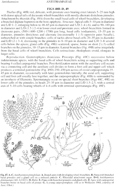
Figure 47 enlarge
Fig. 47. A–C. Antithamnion pinnafolium. A. Branch apex with developing whorl-branchlets. B. Pinna with branched basal pinnules and a gland cell on a reduced pinnule. C. Rhizoidal attachment organ. D–G Antithamnion diminuatum (AD, A13031). D. Habit. E. Pinna with branched pinnules and gland cells. F. Apex with carpogonial branches borne on basal cells of pinnae. G. A young carposporophyte. (All as in Wollaston 1968, courtesy of Aust. J Bot.)
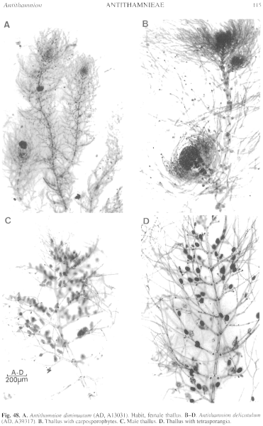
Figure 48 enlarge
Fig. 48. A. Antithamnion diminuatum (AD, A13031). Habit, female thallus. B–D. Antithamnion delicatulum (AD, A39317). B. Thallus with carposporophytes. C. Male thallus. D. Thallus with tetrasporangia.

|
Email Contact: State Herbarium of South Australia |

|


