|
|
|
|
|
|||||||||||
|
Electronic Flora of South Australia Species Fact Sheet
Phylum Rhodophyta – Order Ceramiales – Family Ceramiaceae – Tribe Heterothamnieae
Selected citations: Athanasiadis 1996: 182, fig. 91. Millar & Kraft 1993: 34. Silva et al. 1996: 374.
Synonym
Antithamnion sp. Womersley & Norris 1959: 828.
Thallus (Pl. 1 fig. 1; Fig. 86A, B;) medium red, erect, 0.5–1.5 cm high or in balls 0.5–1 cm across, with subdichotomous axes bearing whorls of 4 (–5) small whorl-branchlets (Fig. 87A) per axial cell. Attachment by branched rhizoids (Figs 86D, 87E) from basal axial cells or from the basal cells of the lowest whorl-branchlets, forming a rhizoidal holdfast; planktonic (free floating) form (Fig. 86B) without attachment; epilithic, epiphytic or free floating. Structure. Apical cells (Fig. 87B, C) tapering, 5–8 µm in basal diameter and L/D 1–1.5, enlarging rapidly (within 4–6 cells) to axial cells 30–50 µm in diameter, then to 120–180 (–220) µm in diameter and L/D 1–2 (–2.5), branched every 2–4 cells (Fig. 87A). Whorl-branchlets unbranched (Fig. 87I) or with 1–3 short branches from the basal cells (Fig. 87D), occasionally digitately branched, 60–90 µm and 4–7 cells long, basal cell largest (20–40 µm in diameter and L/D 0.5–1), then tapering markedly, terminal cell acute, (4–) 6–10 µm in basal diameter and L/D 1–1.5; hairs frequent on any cells of whorl-branchlets; gland cells absent. Lateral branches from just subapical cells, the axes becoming subdichotomous (Fig. 87B), or (especially in female plants) from the basal cells of whorl-branchlets below the procarps. Cells uninucleate; rhodoplasts discoid to elongate in smaller cells, becoming ribbon like in axial cells.
Reproduction: Gametophytes dioecious. Carpogonial branches (Fig. 87F) borne on the basal (= supporting) cell of whorl-branchlets 3–5 cells long, close to apices of axes. Post-
Type from Bridgewater Bay, Vic. (Beauglehole, 21.xi.1961); holotype in AD, A29292.
Selected specimens: Dongara, W. Aust., drift (Wood, 2.x.1960; AD, A24554). Wanna, S. Aust., on Laurencia filiformis, upper sublittoral (Kraft, 11.xi.1971; AD, A42196). Redcliff Point, Spencer Gulf, S. Aust., on Posidonia sinuosa, 7 m deep (Johnson, 26.xi.1975; AD, A50495). West I., S. Aust., on Pterocladia lucida, 18 m deep (Shepherd, 17.viii.1968; AD, A32636). Pennington Bay, Kangaroo I., S. Aust., on Caulerpa, drift (Womersley, 22.viii.1954; AD, A19738) and on Griffithsia monilis, sublittoral fringe (Womersley, 21.xi.1968; AD, A32900). Bridgewater Bay, Vic., free floating (Womersley, 14.iv.1959; AD, A22270 - "Marine Algae of southern Australia" No. 103). Port Fairy, Vic., drift (Fuhrer, 18.ii.1962; AD, A29871). Warrnambool, Vic., on Heterozostera, drift (Womersley, 14.x.1985; AD, A56870). Gabo I., Vic., on Plocamium costatum, 18 m deep (Shepherd, 17.ii.1973; AD, A43510). Fluted Cape, Bruny I., Tas., on Rhodymenia obtusa, 0–6 m deep (Shepherd, 12.ii.1972; AD, A41877).
Distribution: Dongara, W. Aust., to Gabo I., Vic. and SE Tasmania. Twofold Bay, N.S.W. (Millar & Kraft 1993: 34).
Taxonomic notes: fertilization the supporting cell enlarges upwards and cuts off the auxiliary cell, which after fusion with the carpogonium divides to form a foot cell and a central cell which produces a terminal and then lateral gonimolobes (Fig. 87G) 200–300 µm across with ovoid carposporangia 20–35 µm in diameter; the axial cell, residual supporting cell and foot cell fuse, with a broader connection to the central cell (Fig. 87G), Lateral branches from the axial cell below loosely surround the carposporophyte. Spermatangia (Fig. 87H) occur on short special adaxial branches on the basal and lower cells of whorl-branchlets, each branch with 2 or 3 tri- or quadrichotomies.
Tetrasporangia (Fig. 87I) occur on basal cells of whorl-branchlets usually singly, sessile, subspherical to slightly ovoid, 40–50 µm in diameter, decussately to tetrahedrally divided.
Amoenothamnion planktonium occurs on a variety of larger algae and seagrasses, with scattered records along the whole southern Australian coast. The free-floating tetrasporangial form is of frequent occurrence in Bridgewater Bay, Victoria, where it was first observed in 1959 (Womersley & Norris 1959) and has been observed by Mr Bill Steele, a previous proprietor of the Bridgewater Bay Kiosk, on numerous occasions mainly during summer months. This form is also known from Port Fairy and Cape Nelson Bay, Victoria.
References:
ATHANASIADIS, A. (1996). Morphology and classification of the Ceramioideae (Rhodophyta) based on phylogenetic principles. Opera Botanica No. 128, pp. 1–216.
MILLAR, A.J.K. & KRAFT, G.T. (1993). Catalogue of marine and freshwater Red Algae (Rhodophyta) of New South Wales, including Lord Howe Island, South-western Pacific. Aust. Syst. Bot. 6, 1–90.
SILVA, P.C., BASSON, P.W. & MOE, R.L. (1996). Catalogue of the Benthic Marine Algae of the Indian Ocean. (University of California Press: Berkeley, Los Angeles & London.)
WOLLASTON, E.M. (1968).Morphology and taxonomy of southern Australian genera of Crouanieae Schmitz (Ceramiaceae, Rhodophyta). Aust. J. Bot. 16, 217–417.
WOMERSLEY, H.B.S. & NORRIS, R.E. (1959). A free floating marine red alga. Nature, Lond. 184, 828.
The Marine Benthic Flora of Southern Australia Part IIIC complete list of references.
Publication:
Womersley, H.B.S. (24 December, 1998)
The Marine Benthic Flora of Southern Australia
Rhodophyta. Part IIIC. Ceramiales – Ceramiaceae, Dasyaceae
©State Herbarium of South Australia, Government of South Australia
Illustrations in Womersley Part IIIA, 1998: PLATE 1 fig. 1; FIGS 86, 87 A–I.
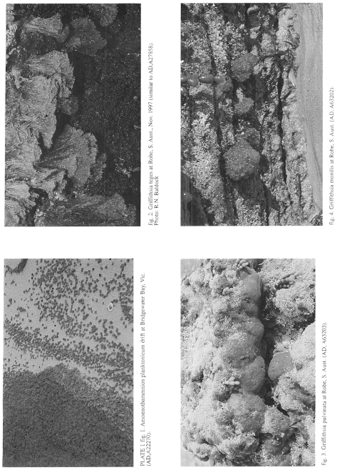
Plate 1 enlarge
PLATE 1 fig. 1. Amoenothamnion planktonium drift at Bridgewater Bay, Vic. (AD, A22270).
fig. 2. Griffithsia teges at Robe, S. Aust., Nov. 1997 (similar to Ad, A27858). Photo: R.N. Baldock.
fig. 3. Griffithsia pulvinata at Robe, S. Aust. (AD, A63203).
fig. 4. Griffithsia monilis at Robe, S. Aust. (AD, A63202)
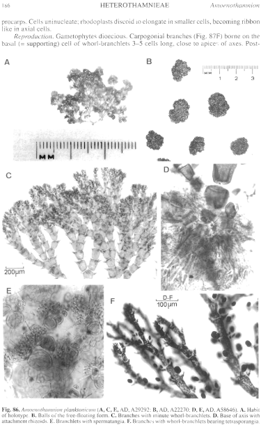
Figure 86 enlarge
Fig. 86. Amoenothamnion planktonicum (A, C, E, AD, A29292; B, AD, A22270; D, E, AD, A58646). A. Habit of holotype. B. Balls of the free-floating form. C. Branches with minute whorl-branchlets. D. Base of axis with attachment rhizoids. E. Branchlets with spermatangia. F. Branches with whorl-branchlets bearing tetrasporangia.
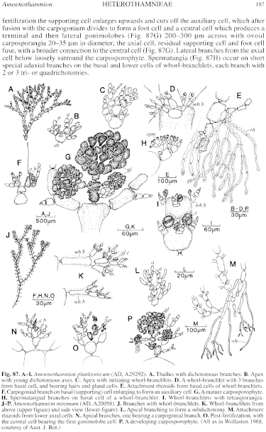
Figure 87 enlarge
Fig. 87.A–I. Amoenothamnion planktonicum (AD, A29292). A. Thallus with dichotomous branches. B. Apex with young dichotomous axes. C. Apex with initiating whorl-branchlets. D. A whorl-branchlet with 3 branches from basal cell, and bearing hairs and gland cells. E. Attachment rhizoids from basal cells of whorl-branchlets. F. Carpogonial branch on basal (supporting) cell enlarging to form an auxiliary cell. G. A mature carposporophyte. H. Spermatangial branches on basal cell of a whorl-branchlet. I. Whorl-branchlets with tetrasporangia. J–P. Amoenothamnion minimum (AD, A20058). J. Branches with whorl-branchlets. K. Whorl-branchlets from above (upper figure) and side view (lower figure). L. Apical branching to form a subdichotomy. M. Attachment rhizoids from lower axial cells. N. Apical branches, one bearing a carpogonial branch. O. Post-fertilization, with the central cell bearing the first gonimolobe cell. P. A developing carposporophyte. (All as in Wollaston 1968. courtesy of Aust. J. Bot.)

|
Email Contact: State Herbarium of South Australia |

|


