|
|
|
|
|
|||||||||||
|
Electronic Flora of South Australia Species Fact Sheet
Phylum Phaeophyta – Order Chordariales – Family Scytothamnaceae
Selected citations: Clayton 1985: 286, figs 1–9. Delépine & Asensi 1978: 129, figs 1,2. Lindauer et al. 1961: 258, fig. 67(1–6). Minter 1984: 87, figs 1–31. Naylor 1955: 295, figs 1–4, pls 6–8. Womersley 1967: 243.
Synonym
A. lessonii (Bory) Hooker & Harvey. Harvey 1858: p1. 48.
Thallus (Fig. 42C) medium to dark brown, with a small discoid holdfast 1–3 mm across bearing one to several clavate to pyriform, clustered, saccate, mucoid vesicles, each with a short solid stipe, (2–) 4–6 (–10) cm long and 0.5–2 (–3) cm in diameter, epilithic or on crustose coralline algae. Structure multiaxial and haplostichous, with an apical pit containing a tuft of phaeophycean hairs (Fig. 431) surrounded by laterally adjacent filaments with periclinal cell divisions, differentiating into a pseudoparenchymatous cortex (Fig. 43J) and loosely filamentous medulla which largely disintegrates leaving the interior mucilage filled. Cortex of 5–8 large cells inwardly, each 25–60 µm in diameter and L/B 1–2, and slightly clavate chains of outer cortical cells, 4–8 cells long, 4–8 µm in diameter with cells L/B (1.5–) 2–3; cortex with numerous, scattered, cryptostomata containing hairs 7–10 µm in diameter. Phaeoplasts discoid, 1–3 per cell, each with a pyrenoid; physodes abundant in outer cortical cells.
Reproduction: Reproduction by unilocular sporangia (Fig. 43J) produced from the subapical cells of some cortical filaments, of which the apical cell extends (L/B 5–10), forming a vague, slightly elevated, sorus developing first on the lower (older) part of the thallus and spreading upwards and extending; sporangia clavate to ovoid, 18–30 µm long and 12–18 µm in diameter.
Type from Iles Malouines (Falkland Is); in Herb. Bory, PC.
Selected specimens: Cape Sorell, Tas., lower eulittoral (Bennett, 4.ii.1955; ADU, A20610). Blow Hole, Eaglehawk Neck, Tas., lower eulittoral on Lithophyllum (Bennett, 8.iii.1958; ADU, A21379). Safety Cove, Port Arthur, Tas., mid eulittoral (Womersley & Parsons, 31.x.1982; ADU, A55951-"Marine Algae of southern Australia" No. 252).
Distribution: Subantarctic islands, Chile, New Zealand (from Wellington south).
In southern Australia, limited to the southern half of Tasmania, and found during summer (October to March) usually in the lower eulittoral on wave-washed rock and in slight pools.
Taxonomic notes: Microthallus gametophytic, filamentous (with some longitudinal divisions in older cells) or discoid, dioecious, producing nodular, plurilocular gametangia with isogametes (which also may come from disc cells), also with parthenogenetic development giving haploid macrothalli (Müller 1984).
Adenocystis is similar in morphology to Utriculidium durvillei (Bory) Skottsberg, from subantarctic islands, the latter bearing only plurilocular organs. While it has been suggested they may be stages of one taxon, Müller (1984) has shown that Adenocystis is a distinct taxon.
References:
CLAYTON, M.N. (1985). A critical investigation of the vegetative anatomy, growth and taxonomic affinities of Adenocystis, Scytothamnus and Splachnidium (Phaeophyta). Br. phycol. 1 20, 285–296.
DELÉPINE, R. & ASENSI, A. (1978). Réactions écophysiologiques et variations morphogénétiques chez Adenocystis et Utriculidium (Phéophycées). Rev. Algol., N.S., 13, 43–85.
HARVEY, W.H. (1858). Phycologia Australica. Vol. I, Plates 1–60. (Reeve: London.)
LINDAUER, V.W., CHAPMAN, V.J. & AIKEN, M. (1961). The marine algae of New Zealand. II. Phaeophyceae. Nova Hedwigia 3, 129–350, Plates 57–97.
MÜLLER, D.G. (1984). Culture studies on the life history of Adenocystis utricularis (Phaeo- . phyceae, Dictyosiphonales). Phycologia 23, 87–94.
NAYLOR, M. (1955). The life history of Adenocystis utricularis (Bory) H. et H. Trans. R. Soc. N.Z. 83, 295–301, Plates 6–8.
SKOTTSBERG, C. (1907). Zur Kenntnis der Subantarktischen und Antarktischen Meeresalgen. I. Phaeophyceen. Wissenschaftliche ergebnisse der Schwedischen Südpolar-expedition 1901–1903, 4, 1–172, Plates 1–10.
SKOTTSBERG, C. (1921). Botanische Ergebnisse der Schwedischen Expedition nach Patagonien und dem Feuerlande, 1907–1909. VIII. Marine Algae. I. Phaeophyceae. K. Svenska Vetenskapsakad. Handl. 61(11), 1–56.
WOMERSLEY, H.B.S. (1967). A critical survey of the marine algae of southern Australia. II. Phaeophyta. Aust. J. Bot. 15, 189–270.
The Marine Benthic Flora of Southern Australia Part II complete list of references.
Publication:
Womersley, H.B.S. (14 December, 1987)
The Marine Benthic Flora of Southern Australia
Part II
©Board of the Botanic Gardens and State Herbarium, Government of South Australia
Illustrations in Womersley Part II, 1997: FIGS 42C, 43I,J.
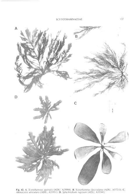
Figure 42 enlarge
Fig. 42. A. Scytothamnus australis (ADU, A29994). B. Scytothamnus fasciculatus (ADU, A57210). C. Adenocystis utricularis (ADU, A55951). D. Splachnidium rugosum (ADU, A55341).
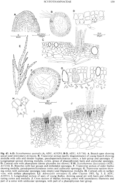
Figure 43 enlarge
Fig. 43. A–D. Scytothamnus australis (A, ADU, A55293; B–D, ADU, A31756). A. Branch apex showing apical (and intercalary) divisions. B. Transverse section (partly diagrammatic) of young branch showing medulla with cells and slender hyphae, pseudoparenchymatous cortex, a hair group and sporangia. C. Longitudinal section showing medulla, cortex, group of phaeophycean hairs and unilocular sporangia. D. Cortical cells with phaeoplasts (dense physodes not shown). E–H. Scytothamnus fasciculatus (ADU, A57210). E. Branches with hair groups and embedded sporangia. F. Transverse section of older thallus showing medulla, pseudoparenchymatous cortex and unilocular sporangia. G. Transverse section showing cortex with unilocular sporangia (one empty) and filamentous medulla. H. Cortical cells in surface view, with stellate phaeoplasts. I,J. Adenocystis utricularis (I, after Clayton 1985, fig. 3; J. ADU, A55951). I. Longitudinal section of apex of young thallus, showing apical pit with hairs and differentiating cortex and medulla. J. Cross section of thallus showing cortex with assimilatory filaments and part of a sorus with unilocular sporangia, with part of a phaeophycean hair group.

|
Email Contact: State Herbarium of South Australia |

|


